

Abstract
OBJECTIVE—To evaluate radiographic features of osteoarthritis (OA) to determine which is more closely associated with knee pain and hence might be used as a radiographic definition of OA in the community. To evaluate joint space width in normal subjects. METHODS—452 subjects from a case-control community study of knee pain (294 women, 158 men, mean age 62 years, range 40-80) underwent AP standing and mid-flexion skyline radiographs. Joint space width, measured by metered calliper to 0.1 mm, and graded individual features of OA (osteophyte 0-3, narrowing 0-3, sclerosis 0-1, cysts 0-1) were assessed in all three compartments independently by two observers who were blind to clinical status. Subjects were categorised as having knee pain by a positive response to both parts of the question "Have you ever had pain in or around the knee on most days for at least a month? If so, have you experienced any pain during the last year?" RESULTS—Intraobserver reproducibility for joint space width measurements was to within ±0.4 mm (95% CI for limits of agreement); κ values for grading were >0.7. One hundred and twenty five subjects were without knee pain or osteophyte. In these radiographically normal knees, mean joint space width varied according to sex but did not decrease with age. A definition based on the presence of osteophyte ⩾grade 1 in any compartment was more efficient at predicting pain than definitions based on either measurement or grading of joint space; there was no clear threshold of joint space loss at which the likelihood of pain substantially increased. The presence of osteophyte at the patellofemoral joint (PFJ) was more sensitive but less specific than at the tibiofemoral joint (TFJ); the addition of PFJ assessment improved sensitivity from 38.1% to 62.3% with a reduction in specificity from 82.7% to 58.7% for the presence of knee pain. CONCLUSION—Among men and women in the community, osteophyte is the radiographic feature that associates best with knee pain. Radiographic assessment of both TFJ and PFJ should be included in all community studies. Joint space loss is not a feature of asymptomatic aging, and there is not a biological cut off for joint space width below which the likelihood of knee pain markedly increases. Keywords: osteoathritis; knee pain
Full Text
The Full Text of this article is available as a PDF (156.7 KB).
Figure 2 .
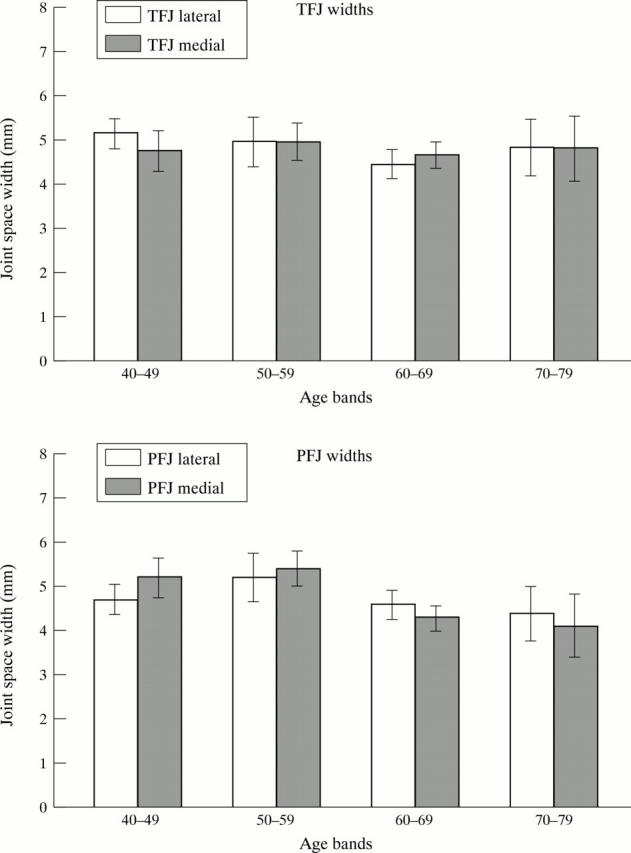
Minimum joint space width (95% CI) in women for lateral and medial TFJ and PFJ compartments.
Figure 1 .
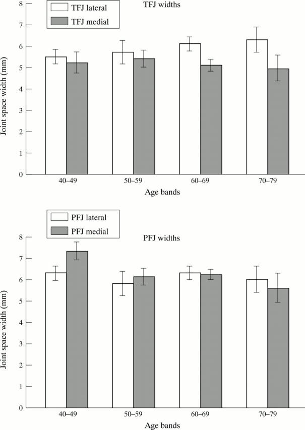
Minimum joint space width (95% CI) in men for medial and lateral TFJ and PFJ compartments.
Selected References
These references are in PubMed. This may not be the complete list of references from this article.
- Altman R. D., Hochberg M., Murphy W. A., Jr, Wolfe F., Lequesne M. Atlas of individual radiographic features in osteoarthritis. Osteoarthritis Cartilage. 1995 Sep;3 (Suppl A):3–70. [PubMed] [Google Scholar]
- Altman R., Asch E., Bloch D., Bole G., Borenstein D., Brandt K., Christy W., Cooke T. D., Greenwald R., Hochberg M. Development of criteria for the classification and reporting of osteoarthritis. Classification of osteoarthritis of the knee. Diagnostic and Therapeutic Criteria Committee of the American Rheumatism Association. Arthritis Rheum. 1986 Aug;29(8):1039–1049. doi: 10.1002/art.1780290816. [DOI] [PubMed] [Google Scholar]
- Bagge E., Bjelle A., Edén S., Svanborg A. Factors associated with radiographic osteoarthritis: results from the population study 70-year-old people in Göteborg. J Rheumatol. 1991 Aug;18(8):1218–1222. [PubMed] [Google Scholar]
- Bland J. M., Altman D. G. Statistical methods for assessing agreement between two methods of clinical measurement. Lancet. 1986 Feb 8;1(8476):307–310. [PubMed] [Google Scholar]
- Buckland-Wright C. Protocols for precise radio-anatomical positioning of the tibiofemoral and patellofemoral compartments of the knee. Osteoarthritis Cartilage. 1995 Sep;3 (Suppl A):71–80. [PubMed] [Google Scholar]
- Buckland-Wright J. C., Macfarlane D. G., Williams S. A., Ward R. J. Accuracy and precision of joint space width measurements in standard and macroradiographs of osteoarthritic knees. Ann Rheum Dis. 1995 Nov;54(11):872–880. doi: 10.1136/ard.54.11.872. [DOI] [PMC free article] [PubMed] [Google Scholar]
- Cicuttini F. M., Baker J., Hart D. J., Spector T. D. Association of pain with radiological changes in different compartments and views of the knee joint. Osteoarthritis Cartilage. 1996 Jun;4(2):143–147. doi: 10.1016/s1063-4584(05)80323-1. [DOI] [PubMed] [Google Scholar]
- Cicuttini F. M., Baker J., Hart D. J., Spector T. D. Choosing the best method for radiological assessment of patellofemoral osteoarthritis. Ann Rheum Dis. 1996 Feb;55(2):134–136. doi: 10.1136/ard.55.2.134. [DOI] [PMC free article] [PubMed] [Google Scholar]
- Croft P., Cooper C., Coggon D. Case definition of hip osteoarthritis in epidemiologic studies. J Rheumatol. 1994 Apr;21(4):591–592. [PubMed] [Google Scholar]
- Croft P., Cooper C., Wickham C., Coggon D. Defining osteoarthritis of the hip for epidemiologic studies. Am J Epidemiol. 1990 Sep;132(3):514–522. doi: 10.1093/oxfordjournals.aje.a115687. [DOI] [PubMed] [Google Scholar]
- DANIELSSON L. G. INCIDENCE AND PROGNOSIS OF COXARTHROSIS. Acta Orthop Scand Suppl. 1964;66:SUPPL 66–7114. doi: 10.3109/ort.1964.35.suppl-66.01. [DOI] [PubMed] [Google Scholar]
- Dacre J. E., Scott D. L., Da Silva J. A., Welsh G., Huskisson E. C. Joint space in radiologically normal knees. Br J Rheumatol. 1991 Dec;30(6):426–428. doi: 10.1093/rheumatology/30.6.426. [DOI] [PubMed] [Google Scholar]
- Danielsson L., Lindberg H., Nilsson B. Prevalence of coxarthrosis. Clin Orthop Relat Res. 1984 Dec;(191):110–115. [PubMed] [Google Scholar]
- Felson D. T., McAlindon T. E., Anderson J. J., Naimark A., Weissman B. W., Aliabadi P., Evans S., Levy D., LaValley M. P. Defining radiographic osteoarthritis for the whole knee. Osteoarthritis Cartilage. 1997 Jul;5(4):241–250. doi: 10.1016/s1063-4584(97)80020-9. [DOI] [PubMed] [Google Scholar]
- Hall M. G., Ferrell W. R., Sturrock R. D., Hamblen D. L., Baxendale R. H. The effect of the hypermobility syndrome on knee joint proprioception. Br J Rheumatol. 1995 Feb;34(2):121–125. doi: 10.1093/rheumatology/34.2.121. [DOI] [PubMed] [Google Scholar]
- Jones A. C., Ledingham J., McAlindon T., Regan M., Hart D., MacMillan P. J., Doherty M. Radiographic assessment of patellofemoral osteoarthritis. Ann Rheum Dis. 1993 Sep;52(9):655–658. doi: 10.1136/ard.52.9.655. [DOI] [PMC free article] [PubMed] [Google Scholar]
- Jones A., Hopkinson N., Pattrick M., Berman P., Doherty M. Evaluation of a method for clinically assessing osteoarthritis of the knee. Ann Rheum Dis. 1992 Feb;51(2):243–245. doi: 10.1136/ard.51.2.243. [DOI] [PMC free article] [PubMed] [Google Scholar]
- Jørring K. Osteoarthritis of the hip. Epidemiology and clinical role. Acta Orthop Scand. 1980 Jun;51(3):523–530. doi: 10.3109/17453678008990835. [DOI] [PubMed] [Google Scholar]
- Lanyon P., Jones A., Doherty M. Assessing progression of patellofemoral osteoarthritis: a comparison between two radiographic methods. Ann Rheum Dis. 1996 Dec;55(12):875–879. doi: 10.1136/ard.55.12.875. [DOI] [PMC free article] [PubMed] [Google Scholar]
- Laurin C. A., Dussault R., Levesque H. P. The tangential x-ray investigation of the patellofemoral joint: x-ray technique, diagnostic criteria and their interpretation. Clin Orthop Relat Res. 1979 Oct;(144):16–26. [PubMed] [Google Scholar]
- McAlindon T. E., Snow S., Cooper C., Dieppe P. A. Radiographic patterns of osteoarthritis of the knee joint in the community: the importance of the patellofemoral joint. Ann Rheum Dis. 1992 Jul;51(7):844–849. doi: 10.1136/ard.51.7.844. [DOI] [PMC free article] [PubMed] [Google Scholar]
- Nevitt M. C. Definition of hip osteoarthritis for epidemiological studies. Ann Rheum Dis. 1996 Sep;55(9):652–655. doi: 10.1136/ard.55.9.652. [DOI] [PMC free article] [PubMed] [Google Scholar]
- O'Reilly S. C., Muir K. R., Doherty M. Screening for pain in knee osteoarthritis: which question? Ann Rheum Dis. 1996 Dec;55(12):931–933. doi: 10.1136/ard.55.12.931. [DOI] [PMC free article] [PubMed] [Google Scholar]
- Petersson I. F. Occurrence of osteoarthritis of the peripheral joints in European populations. Ann Rheum Dis. 1996 Sep;55(9):659–661. doi: 10.1136/ard.55.9.659. [DOI] [PMC free article] [PubMed] [Google Scholar]
- Spector T. D., Hart D. J., Byrne J., Harris P. A., Dacre J. E., Doyle D. V. Definition of osteoarthritis of the knee for epidemiological studies. Ann Rheum Dis. 1993 Nov;52(11):790–794. doi: 10.1136/ard.52.11.790. [DOI] [PMC free article] [PubMed] [Google Scholar]
- Spector T. D., Hart D. J. How serious is knee osteoarthritis? Ann Rheum Dis. 1992 Oct;51(10):1105–1106. doi: 10.1136/ard.51.10.1105. [DOI] [PMC free article] [PubMed] [Google Scholar]