

Full Text
The Full Text of this article is available as a PDF (121.8 KB).
Figure 1 .
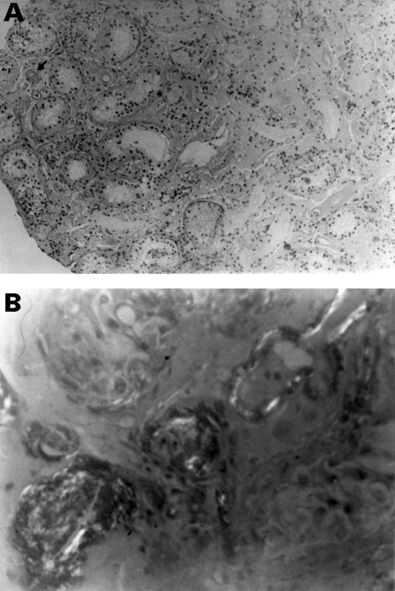
(A) Testicular biopsy specimen obtained from patient A, showing atrophy and fibrosis of the tubules without Leydig cell hyperplasia. Note the thickening of the walls of the tubules and the blood vessels (arrow). (B) Illustrating birefringence of amyloid deposits in tubular and blood vessels walls after Congo red staining.
Figure 2 .
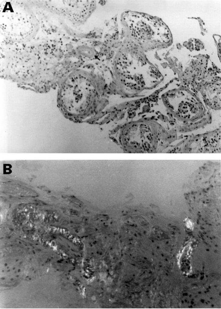
(A) Testicular biopsy specimen obtained from patient C, disclosed maturation arrest and thickening of the walls of the blood vessels (arrow). (B) Illustrating birefringence of amyloid deposits in tubules and blood vessels after Congo red staining.