Abstract
OBJECTIVE—To compare increased bone uptake of 99Tcm-MDP and magnetic resonance (MR) detected subchondral lesions, osteophytes, and cartilage defects in the knee in middle aged people with longstanding knee pain. METHODS—Fifty eight people (aged 41-58 years, mean 50) with chronic knee pain, with or without radiographic knee osteoarthritis, were examined with bone scintigraphy. The pattern and the grade of increased bone uptake was assessed. On the same day, a MR examination on a 1.0 T imager was performed. The presence and the grade of subchondral lesions, osteophytes, and cartilage defects were registered. RESULTS—The κ values describing the correlation between increased bone uptake and MR detected subchondral lesions varied between 0.79 and 0.49, and between increased bone uptake and MR detected osteophytes or cartilage defects the values were <0.54. The κ values describing the correlation between the grade of bone uptake and the grade of the different MR findings was <0.57. CONCLUSIONS—Good agreement was found between increased bone uptake and MR detected subchondral lesion. The agreement between increased bone uptake and osteophytes or cartilage defects was in general poor as well as the agreement between the grade of bone uptake and the grade of the MR findings. Keywords: knee; osteoarthritis; magnetic resonance imaging; bone scintigraphy
Full Text
The Full Text of this article is available as a PDF (184.3 KB).
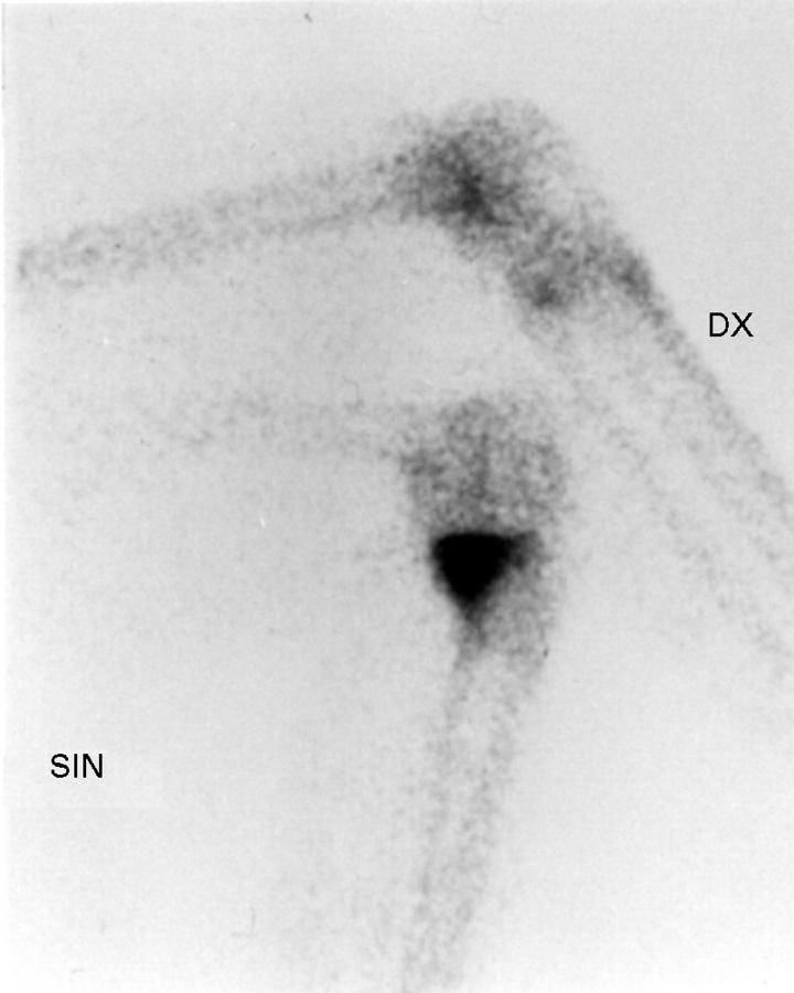
Figure 1 .
Bone scintigram of the knees in right medial and left lateral projection in a 51 year old woman, showing an increased bone uptake with a point-like pattern of grade 2 in the medial femoral condyle of the right knee (the signal knee). The left knee has normal uptake.
Figure 2 .
Bone scintigram of the knees in right medial and left lateral projection in a 58 year old woman, demonstrating an increased bone uptake with a tramline pattern of grade 1 in the lateral tibial condyle of the left knee (arrow) (the signal knee) as well as in the medial tibial condyle of the right knee (double arrow).
Figure 3 .
Bone scintigram of the knees in right lateral and left medial projections in a 42 year old woman, showing an increased bone uptake with an extended pattern in the medial tibial condyle of the left knee (the signal knee). The right knee has normal uptake.
Figure 4 .

(A) A sagittal T2 weighted STIR+ MR image of the right knee in a 43 year old man demonstrating a subchondral lesion of grade 2 dorsal in the lateral femoral condyle as well as in the lateral tibial condyle (arrows). (B) In a corresponding sagittal proton density weighted MR image is shown adjacent cartilage defects of grade 2 in both locations (arrows).
Figure 5 .
A coronal proton density weighted MR image of the right knee in a 57 year old woman demonstrating a peripheral osteophyte of grade 1, at the medial femoral condyle (white arrow). Note the cartilage defect of grade 2 in the same condyle (black arrow).
Selected References
These references are in PubMed. This may not be the complete list of references from this article.
- Bagge E., Bjelle A., Svanborg A. Radiographic osteoarthritis in the elderly. A cohort comparison and a longitudinal study of the "70-year old people in Göteborg". Clin Rheumatol. 1992 Dec;11(4):486–491. doi: 10.1007/BF02283103. [DOI] [PubMed] [Google Scholar]
- Boegård T., Rudling O., Petersson I. F., Sanfridsson J., Saxne T., Svensson B., Jonsson K. Joint-space width in the axial view of the patello-femoral joint. Definitions and comparison with MR imaging. Acta Radiol. 1998 Jan;39(1):24–31. doi: 10.1080/02841859809172144. [DOI] [PubMed] [Google Scholar]
- Boegård T., Rudling O., Petersson I. F., Sanfridsson J., Saxne T., Svensson B., Jonsson K. Postero-anterior radiogram of the knee in weight-bearing and semiflexion. Comparison with MR imaging. Acta Radiol. 1997 Nov;38(6):1063–1070. doi: 10.1080/02841859709172132. [DOI] [PubMed] [Google Scholar]
- Broderick L. S., Turner D. A., Renfrew D. L., Schnitzer T. J., Huff J. P., Harris C. Severity of articular cartilage abnormality in patients with osteoarthritis: evaluation with fast spin-echo MR vs arthroscopy. AJR Am J Roentgenol. 1994 Jan;162(1):99–103. doi: 10.2214/ajr.162.1.8273700. [DOI] [PubMed] [Google Scholar]
- Brown T. R., Quinn S. F. Evaluation of chondromalacia of the patellofemoral compartment with axial magnetic resonance imaging. Skeletal Radiol. 1993;22(5):325–328. doi: 10.1007/BF00198391. [DOI] [PubMed] [Google Scholar]
- Christensen S. B. Localization of bone-seeking agents in developing, experimentally induced osteoarthritis in the knee joint of the rabbit. Scand J Rheumatol. 1983;12(4):343–349. doi: 10.3109/03009748309099738. [DOI] [PubMed] [Google Scholar]
- Cicuttini F. M., Spector T., Baker J. Risk factors for osteoarthritis in the tibiofemoral and patellofemoral joints of the knee. J Rheumatol. 1997 Jun;24(6):1164–1167. [PubMed] [Google Scholar]
- Dieppe P., Cushnaghan J., Young P., Kirwan J. Prediction of the progression of joint space narrowing in osteoarthritis of the knee by bone scintigraphy. Ann Rheum Dis. 1993 Aug;52(8):557–563. doi: 10.1136/ard.52.8.557. [DOI] [PMC free article] [PubMed] [Google Scholar]
- Dieppe P. Osteoarthritis and molecular markers. A rheumatologist's perspective. Acta Orthop Scand Suppl. 1995 Oct;266:1–5. [PubMed] [Google Scholar]
- Dieppe P. Osteoarthritis: clinical and research perspective. Br J Rheumatol. 1991;30 (Suppl 1):1–4. [PubMed] [Google Scholar]
- Disler D. G. Fat-suppressed three-dimensional spoiled gradient-recalled MR imaging: assessment of articular and physeal hyaline cartilage. AJR Am J Roentgenol. 1997 Oct;169(4):1117–1123. doi: 10.2214/ajr.169.4.9308475. [DOI] [PubMed] [Google Scholar]
- Dye S. F., Boll D. A. Radionuclide imaging of the patellofemoral joint in young adults with anterior knee pain. Orthop Clin North Am. 1986 Apr;17(2):249–262. [PubMed] [Google Scholar]
- Egund N., Frost S., Brismar J., Gustafson T. Radiography and scintigraphy in the assessment of early gonarthrosis. Acta Radiol. 1988 Jul-Aug;29(4):451–455. [PubMed] [Google Scholar]
- Gilbertson E. M. Development of periarticular osteophytes in experimentally induced osteoarthritis in the dog. A study using microradiographic, microangiographic, and fluorescent bone-labelling techniques. Ann Rheum Dis. 1975 Feb;34(1):12–25. doi: 10.1136/ard.34.1.12. [DOI] [PMC free article] [PubMed] [Google Scholar]
- Gross W. L., Lohmann C., Lúdemann G., Lúdemann J. Lymphocyte response to enterobacterial biostructures in seronegative spondarthritis: specific T-cell mediated immunity or non-specific polyclonal B-cell activation? Br J Rheumatol. 1988;27 (Suppl 2):23–28. doi: 10.1093/rheumatology/xxvii.suppl_2.23. [DOI] [PubMed] [Google Scholar]
- Hayes C. W., Sawyer R. W., Conway W. F. Patellar cartilage lesions: in vitro detection and staging with MR imaging and pathologic correlation. Radiology. 1990 Aug;176(2):479–483. doi: 10.1148/radiology.176.2.2367664. [DOI] [PubMed] [Google Scholar]
- Hernborg J., Nilsson B. E. The relationship between osteophytes in the knee joint, osteoarthritis and aging. Acta Orthop Scand. 1973;44(1):69–74. doi: 10.3109/17453677308988675. [DOI] [PubMed] [Google Scholar]
- Heron C. W., Calvert P. T. Three-dimensional gradient-echo MR imaging of the knee: comparison with arthroscopy in 100 patients. Radiology. 1992 Jun;183(3):839–844. doi: 10.1148/radiology.183.3.1584944. [DOI] [PubMed] [Google Scholar]
- Hilfiker P., Zanetti M., Debatin J. F., McKinnon G., Hodler J. Fast spin-echo inversion-recovery imaging versus fast T2-weighted spin-echo imaging in bone marrow abnormalities. Invest Radiol. 1995 Feb;30(2):110–114. doi: 10.1097/00004424-199502000-00009. [DOI] [PubMed] [Google Scholar]
- Hutton C. W., Higgs E. R., Jackson P. C., Watt I., Dieppe P. A. 99mTc HMDP bone scanning in generalised nodal osteoarthritis. I. Comparison of the standard radiograph and four hour bone scan image of the hand. Ann Rheum Dis. 1986 Aug;45(8):617–621. doi: 10.1136/ard.45.8.617. [DOI] [PMC free article] [PubMed] [Google Scholar]
- Hutton C. W., Higgs E. R., Jackson P. C., Watt I., Dieppe P. A. 99mTc HMDP bone scanning in generalised nodal osteoarthritis. II. The four hour bone scan image predicts radiographic change. Ann Rheum Dis. 1986 Aug;45(8):622–626. doi: 10.1136/ard.45.8.622. [DOI] [PMC free article] [PubMed] [Google Scholar]
- Karvonen R. L., Negendank W. G., Fraser S. M., Mayes M. D., An T., Fernandez-Madrid F. Articular cartilage defects of the knee: correlation between magnetic resonance imaging and gross pathology. Ann Rheum Dis. 1990 Sep;49(9):672–675. doi: 10.1136/ard.49.9.672. [DOI] [PMC free article] [PubMed] [Google Scholar]
- McAlindon T. E., Snow S., Cooper C., Dieppe P. A. Radiographic patterns of osteoarthritis of the knee joint in the community: the importance of the patellofemoral joint. Ann Rheum Dis. 1992 Jul;51(7):844–849. doi: 10.1136/ard.51.7.844. [DOI] [PMC free article] [PubMed] [Google Scholar]
- McAlindon T. E., Watt I., McCrae F., Goddard P., Dieppe P. A. Magnetic resonance imaging in osteoarthritis of the knee: correlation with radiographic and scintigraphic findings. Ann Rheum Dis. 1991 Jan;50(1):14–19. doi: 10.1136/ard.50.1.14. [DOI] [PMC free article] [PubMed] [Google Scholar]
- McCrae F., Shouls J., Dieppe P., Watt I. Scintigraphic assessment of osteoarthritis of the knee joint. Ann Rheum Dis. 1992 Aug;51(8):938–942. doi: 10.1136/ard.51.8.938. [DOI] [PMC free article] [PubMed] [Google Scholar]
- McDevitt C., Gilbertson E., Muir H. An experimental model of osteoarthritis; early morphological and biochemical changes. J Bone Joint Surg Br. 1977 Feb;59(1):24–35. doi: 10.1302/0301-620X.59B1.576611. [DOI] [PubMed] [Google Scholar]
- Mirowitz S. A., Apicella P., Reinus W. R., Hammerman A. M. MR imaging of bone marrow lesions: relative conspicuousness on T1-weighted, fat-suppressed T2-weighted, and STIR images. AJR Am J Roentgenol. 1994 Jan;162(1):215–221. doi: 10.2214/ajr.162.1.8273669. [DOI] [PubMed] [Google Scholar]
- Peterfy C. G. MR imaging. Baillieres Clin Rheumatol. 1996 Nov;10(4):635–678. doi: 10.1016/s0950-3579(96)80055-0. [DOI] [PubMed] [Google Scholar]
- Peterfy C. G., van Dijke C. F., Janzen D. L., Glüer C. C., Namba R., Majumdar S., Lang P., Genant H. K. Quantification of articular cartilage in the knee with pulsed saturation transfer subtraction and fat-suppressed MR imaging: optimization and validation. Radiology. 1994 Aug;192(2):485–491. doi: 10.1148/radiology.192.2.8029420. [DOI] [PubMed] [Google Scholar]
- Petersson I. F., Boegård T., Dahlström J., Svensson B., Heinegård D., Saxne T. Bone scan and serum markers of bone and cartilage in patients with knee pain and osteoarthritis. Osteoarthritis Cartilage. 1998 Jan;6(1):33–39. doi: 10.1053/joca.1997.0090. [DOI] [PubMed] [Google Scholar]
- Petersson I. F., Boegård T., Saxne T., Silman A. J., Svensson B. Radiographic osteoarthritis of the knee classified by the Ahlbäck and Kellgren & Lawrence systems for the tibiofemoral joint in people aged 35-54 years with chronic knee pain. Ann Rheum Dis. 1997 Aug;56(8):493–496. doi: 10.1136/ard.56.8.493. [DOI] [PMC free article] [PubMed] [Google Scholar]
- Recht M. P., Piraino D. W., Paletta G. A., Schils J. P., Belhobek G. H. Accuracy of fat-suppressed three-dimensional spoiled gradient-echo FLASH MR imaging in the detection of patellofemoral articular cartilage abnormalities. Radiology. 1996 Jan;198(1):209–212. doi: 10.1148/radiology.198.1.8539380. [DOI] [PubMed] [Google Scholar]
- Smith R. C., Constable R. T., Reinhold C., McCauley T., Lange R. C., McCarthy S. Fast spin echo STIR imaging. J Comput Assist Tomogr. 1994 Mar-Apr;18(2):209–213. doi: 10.1097/00004728-199403000-00007. [DOI] [PubMed] [Google Scholar]
- Tervonen O., Dietz M. J., Carmichael S. W., Ehman R. L. MR imaging of knee hyaline cartilage: evaluation of two- and three-dimensional sequences. J Magn Reson Imaging. 1993 Jul-Aug;3(4):663–668. doi: 10.1002/jmri.1880030417. [DOI] [PubMed] [Google Scholar]
- Thomas R. H., Resnick D., Alazraki N. P., Daniel D., Greenfield R. Compartmental evaluation of osteoarthritis of the knee. A comparative study of available diagnostic modalities. Radiology. 1975 Sep;116(3):585–594. doi: 10.1148/116.3.585. [DOI] [PubMed] [Google Scholar]


