Abstract
OBJECTIVE—The aim of this pictorial essay is to describe the sonographic guided approach to investigation and local injection therapy of a small joint in a patient with psoriatic arthritis (PA). METHODS—Sonographic pictures are obtained using a high frequency ultrasonography apparatus equipped with a 13-MHz transducer. RESULTS—Ultrasonography allows a careful morphostructural assessment of soft tissue involvement in PA patients. Sonographic findings include joint cavity widening, capsular thickening, synovial proliferation, synovial fluid changes, tendon sheath widening. Ultrasound guided placement of the needle within the joint and injection of corticosteroid under sonographic control can be easily performed. CONCLUSIONS—High frequency ultrasonography is a quick and safe procedure that allows a useful diagnostic and therapeutic approach in patients with arthritis of small joints.
Full Text
The Full Text of this article is available as a PDF (2.4 MB).
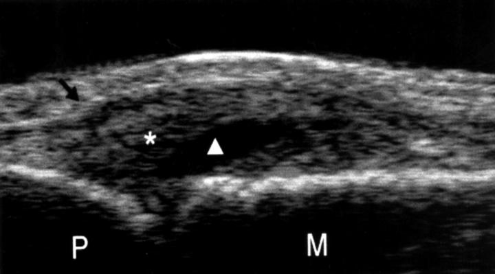
Figure 1 .
Longitudinal dorsal scan showing a clearly evident space widening of the MCP joint, synovial fluid, and synovial proliferation. Black arrow = extensor tendon; * = synovial proliferation; white triangle = synovial fluid; P =proximal phalanx; M = metacarpal bone.
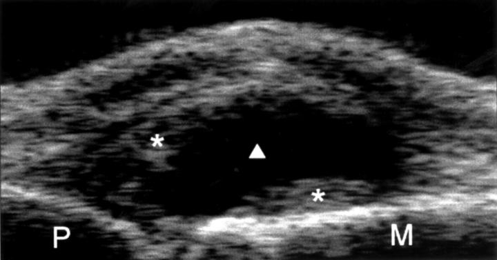
Figure 2 .
Longitudinal dorsal scan with the finger in passive hyperextension confirms that the mildly echogenic material within the joint space (*) can be regarded as pannus. White triangle = synovial fluid; P = proximal phalanx; M = metacarpal bone.
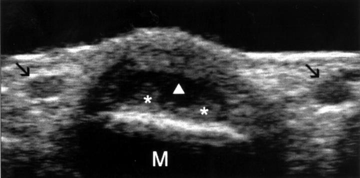
Figure 3 .
Transverse dorsal scan. Any doubt concerning the presence of proliferative synovitis is removed by a dorsal transverse view showing two demarcated areas of synovial hypertrophy(*). Black arrow = common palmar digital artery; white triangle = synovial fluid; M = metacarpal bone.
Figure 4 .
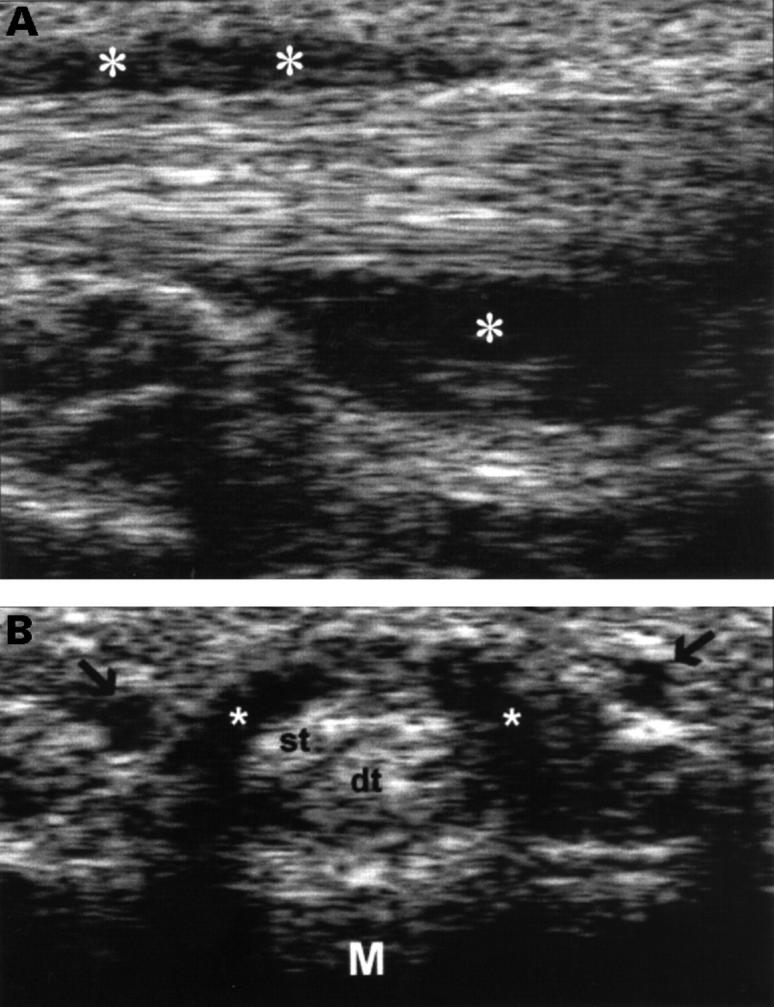
(A) Longitudinal sonogram of the MCP joint along the finger flexor tendons. This view allows identification of a clearly evident tendon sheath widening (*). (B) Transverse sonogram of the same area confirms the presence of a homogeneous anechoic widening of the tendon sheath (*). M = metacarpal bone; st = superficial flexor tendon; dt = deep flexor tendon; black arrows = common palmar digital artery.
Figure 5 .
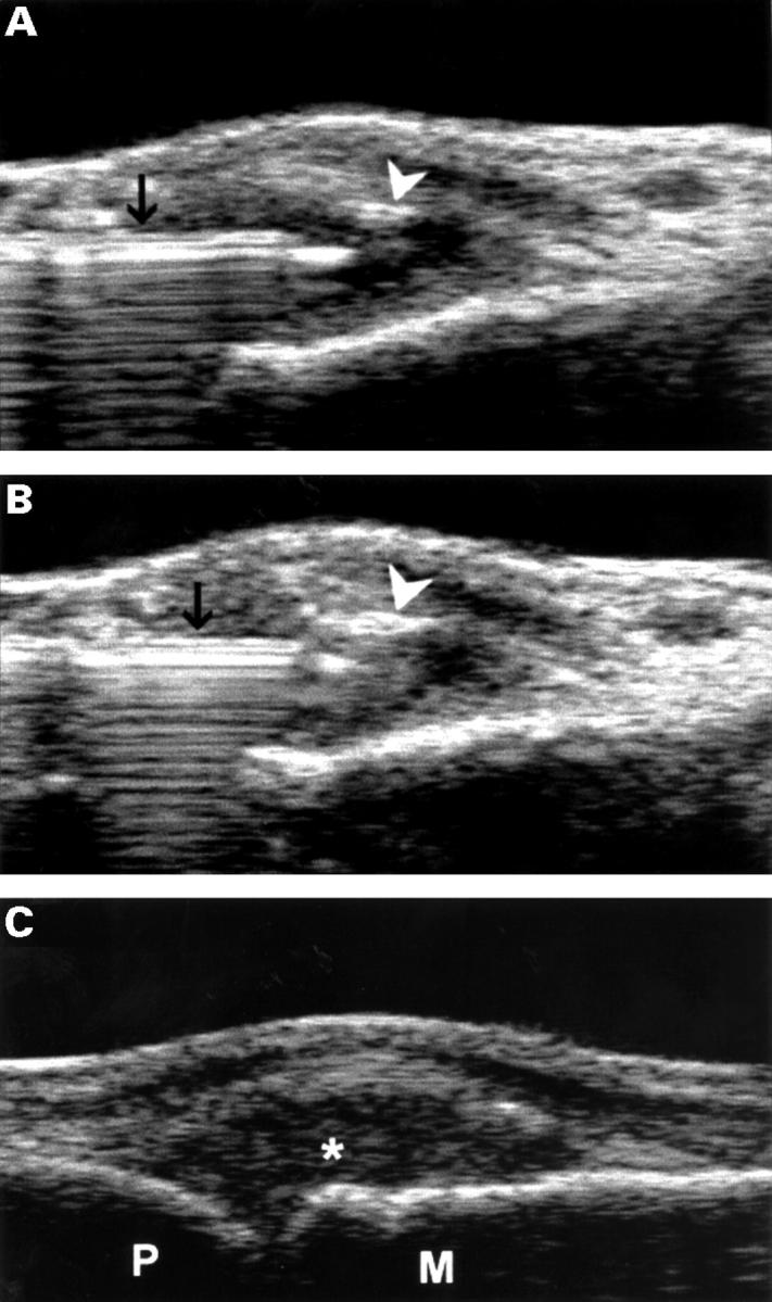
(A) Longitudinal sonogram of the MCP joint showing the placement of the tip of the needle within the joint cavity. The image has been obtained after the injection with a minimal amount of triamcinolone hexacetonide. Triamcinolone drops are highly echoic and can be easily visualised as white spots (arrowhead). Black arrow = needle. (B) Complete filling of the joint cavity by corticosteroid. (C) Longitudinal sonogram of the MCP joint after the injection shows an echoic joint cavity (*). M = metacarpal bone; P = proximal phalanx.
Selected References
These references are in PubMed. This may not be the complete list of references from this article.
- Grassi W., Cervini C. Ultrasonography in rheumatology: an evolving technique. Ann Rheum Dis. 1998 May;57(5):268–271. doi: 10.1136/ard.57.5.268. [DOI] [PMC free article] [PubMed] [Google Scholar]
- Grassi W., Tittarelli E., Blasetti P., Pirani O., Cervini C. Finger tendon involvement in rheumatoid arthritis. Evaluation with high-frequency sonography. Arthritis Rheum. 1995 Jun;38(6):786–794. doi: 10.1002/art.1780380611. [DOI] [PubMed] [Google Scholar]
- Grassi W., Tittarelli E., Pirani O., Avaltroni D., Cervini C. Ultrasound examination of metacarpophalangeal joints in rheumatoid arthritis. Scand J Rheumatol. 1993;22(5):243–247. doi: 10.3109/03009749309095131. [DOI] [PubMed] [Google Scholar]
- Lehtinen A., Taavitsainen M., Leirisalo-Repo M. Sonographic analysis of enthesopathy in the lower extremities of patients with spondylarthropathy. Clin Exp Rheumatol. 1994 Mar-Apr;12(2):143–148. [PubMed] [Google Scholar]
- Manger B., Kalden J. R. Joint and connective tissue ultrasonography--a rheumatologic bedside procedure? A German experience. Arthritis Rheum. 1995 Jun;38(6):736–742. doi: 10.1002/art.1780380603. [DOI] [PubMed] [Google Scholar]
- Martinoli C., Derchi L. E., Pastorino C., Bertolotto M., Silvestri E. Analysis of echotexture of tendons with US. Radiology. 1993 Mar;186(3):839–843. doi: 10.1148/radiology.186.3.8430196. [DOI] [PubMed] [Google Scholar]


