

Abstract
OBJECTIVE—To report the efficacy of conservative treatment with cervical traction and immobilisation with a Halo vest, in two consecutive rheumatoid arthritis patients with progressive cervical myelopathy caused by subaxial subluxation. METHODS—Description of neurological symptoms and signs and findings in plain radiography (PR) and magnetic resonance imaging (MRI) of the cervical spine before and after treatment of the subaxial subluxation by traction and immobilisation with a Halo vest during four months. RESULTS—During four months of traction and immobilisation neurological examination showed a considerable improvement of the signs and symptoms of cervical myelopathy. Afterwards PR and MRI of the cervical spine showed reduction of the subaxial subluxation. Eventually firm stabilisation was obtained in both patients without surgery of the cervical spine. CONCLUSION—Cervical traction and immobilisation with a Halo vest can be considered as an independent conservative treatment in rheumatoid arthritis patients with cervical myelopathy caused by subaxial subluxation. Keywords: rheumatoid arthrits; rheumatoid subaxial subluxation
Full Text
The Full Text of this article is available as a PDF (147.8 KB).
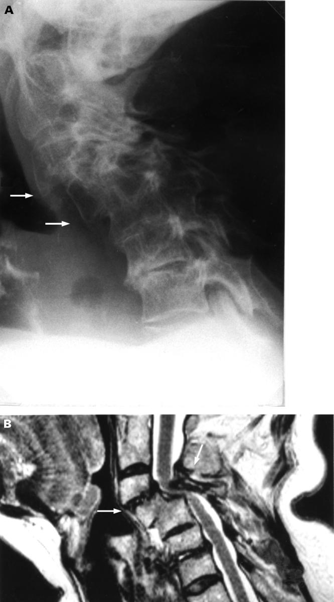
Figure 1 .

Lateral plain radiograph in flexion (A) and T2 weighted sagittal MR image (B) of the cervical spine showing C4-C5 subluxation of 9 mm with secondary spondylosis, discopathy, narrowing of the spinal subarachnoid space, and compression of the spinal cord.
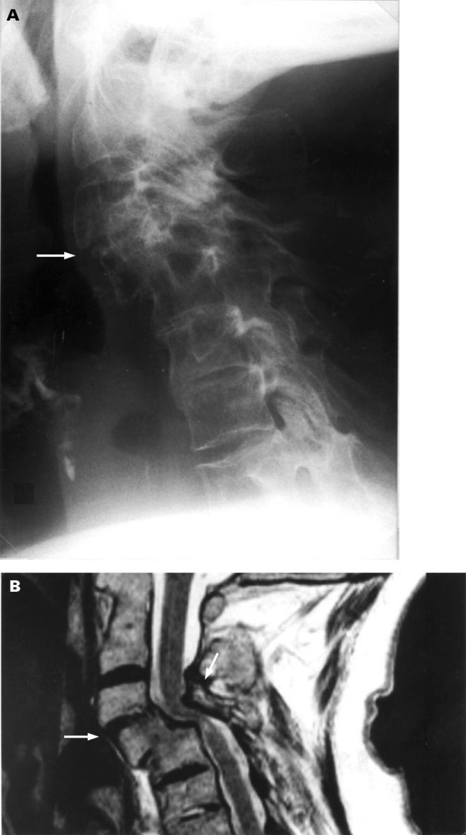
Figure 2 .

Lateral plain radiography (A) and T2 weighted sagittal MR image (B) of the cervical spine after four months Halo vest traction and immobilisation showing reduction of C4-C5 subluxation, narrowing of the spinal subarachnoid space, and compression of the spinal cord.
Figure 3 .
Lateral plain radiography in flexion (A) and T2 weighted sagittal MR image (B) of the cervical spine showing C3-C4 subluxation of 9 mm, stable C4-C5 subluxation of 8 mm with fusion of the vertebral bodies of C4 and C5, narrowing of the spinal subarachnoid space, and compression of the spinal cord.
Figure 4 .
Lateral plain radiography (A) and T2 weighted sagittal MR image (B) of the cervical spine showing reduction and stabilisation of C3-C4 subluxation (3 mm), reduction of compression of the spinal cord, and no changes at C4-C5 level.