Abstract
OBJECTIVES—To examine the expression of the p53 protein in synovial membrane of rheumatoid arthritis (RA) patients and to compare this with the expression in normal synovial tissues in subjects without RA. METHODS—Immunohistological expression of the p53 protein was studied using a streptavidin-biotin-peroxidase method and the monoclonal antibody DO-7, an antibody directed against both wild and mutant forms of p53 protein, in synovial tissues of RA patients (n=10) and from subjects with no known joint disease (n=4). RESULTS—p53 protein expression was present in a small percentage of synovial cells in the majority of the RA patients (n=8; 80%) and in half of the normal control cases with no inflammatory joint disease (n=2; 50%). No sample had more than 5% cells staining with intranuclear pattern. The difference in synovial p53 immunoreactivity between the RA patients and normal controls is not statistically significant (p= 0.64; χ2 contingency test). CONCLUSIONS—This study has shown that p53 protein is only weakly expressed in the rheumatoid synovial membrane, with a low percentage of p53 protein immunostaining cells present, with intranuclear staining. These results suggest this is wild type p53 protein rather than mutant protein. These findings suggest that synovial p53 protein expression may not be important in the pathogenesis of RA and may only represent a reactive repair process to DNA damage secondary to the immune and inflammatory reactions associated with the disease.
Full Text
The Full Text of this article is available as a PDF (132.5 KB).
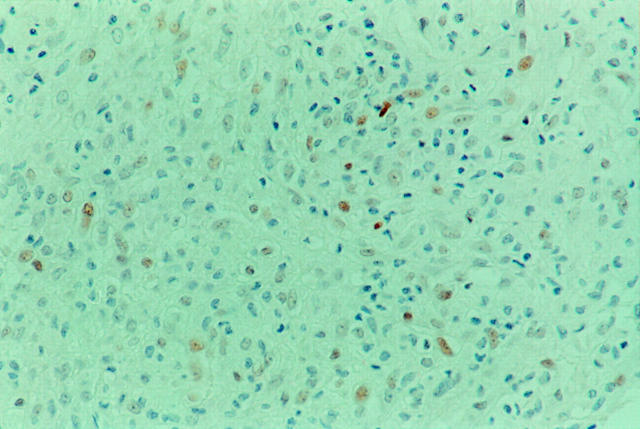
Figure 1 .
p53 protein nuclear immunostaining present in a small number of synovial cells in a case of clinically active rheumatoid arthritis. (Immunoperoxidase, DO-7; original magnification × 200).
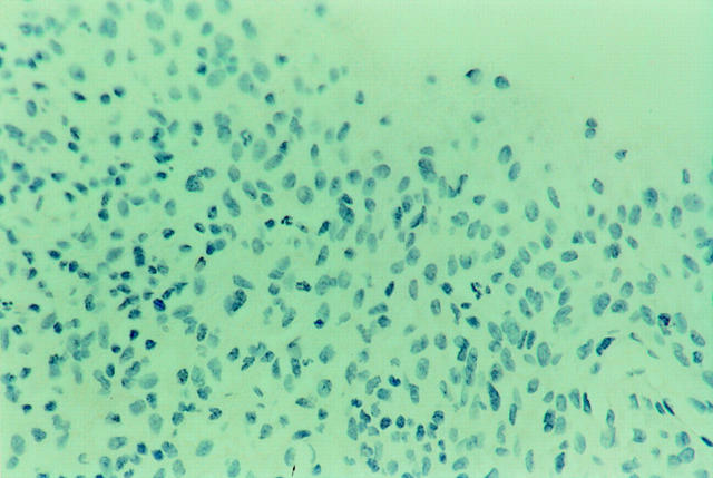
Figure 2 .
Absence of p53 protein immunostaining in the normal synovium of a subject with no inflammatory joint disease. (Immunoperoxidase, DO-7; original magnification × 200).


