

Abstract
OBJECTIVES—To examine objectively spatial patterns of osteophytes around the distal end of the femur and to identify distinct subgroups. METHODS—A sample of 107 human femora from a large skeletal population were selected for study. These femora included subjects with evidence of late stage osteoarthritis (that is, with eburnation present) and those with no such evidence. The location of osteophytes was recorded using a video camera and digitised computer images were extracted. Multidimensional scaling was used to identify clusters of femora based upon osteophyte location. RESULTS—A distinct subgroup of femora was identified with osteophytes present only within the intercondylar notch region. None of these subjects had any evidence of eburnation. CONCLUSIONS—This finding adds to an earlier study based on radiographs. Osteophytes located within the intercondylar notch of the femur appear to be a distinct subset, which may occur either as an early stage of knee osteoarthritis or for some independent reason.
Full Text
The Full Text of this article is available as a PDF (341.4 KB).
Figure 1 .

Example of colour coded bitmap. Red = osteophytes; blue = eburnation; yellow = damage.
Figure 2 .

Configuration of femora in three dimensions. Smaller crosses indicate higher scores in all three dimensions (that is, further from the "viewpoint").
Figure 3 .
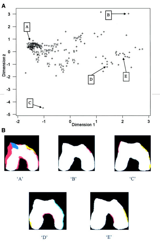
(A) Configuration of femora with osteophytes in dimensions 1 and 2—all osteophytic femora. PL = lateral patellofemoral eburnation; PM = medial patellofemoral eburnation; TM = medial tibiofemoral eburnation. (B) Examples of femora from regions `A' to `E' identified from the multidimensional scaling—configuration projected into dimensions 1 and 2.
Figure 4 .
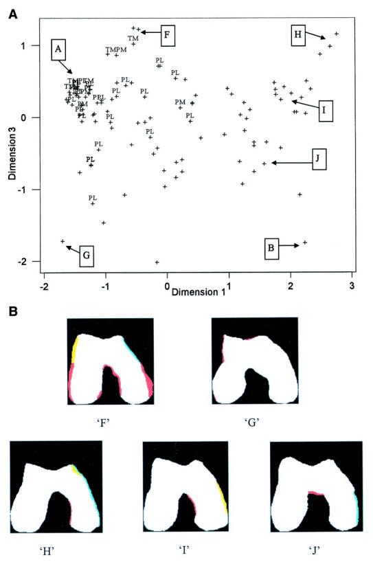
(A) Configuration of femora with osteophytes in dimensions 1 and 3— all osteophytic femora. PL = lateral patellofemoral eburnation; PM = medial patellofemoral eburnation; TM = medial tibiofemoral eburnation. (B) Examples of femora from regions `F' to `J' identified from the multidimensional scaling—configuration projected into dimensions 1 and 3.
Figure 5 .

Configuration of femora with osteophytes in dimensions 1 and 2 (all osteophytic femora) with indication of cluster membership. Different symbols indicate four different clusters. Femora identified with `x' have lone intercondylar notch osteophytes and form a distinct cluster from the cluster analysis.
Selected References
These references are in PubMed. This may not be the complete list of references from this article.
- Altman R., Asch E., Bloch D., Bole G., Borenstein D., Brandt K., Christy W., Cooke T. D., Greenwald R., Hochberg M. Development of criteria for the classification and reporting of osteoarthritis. Classification of osteoarthritis of the knee. Diagnostic and Therapeutic Criteria Committee of the American Rheumatism Association. Arthritis Rheum. 1986 Aug;29(8):1039–1049. doi: 10.1002/art.1780290816. [DOI] [PubMed] [Google Scholar]
- Cicuttini F. M., Spector T., Baker J. Risk factors for osteoarthritis in the tibiofemoral and patellofemoral joints of the knee. J Rheumatol. 1997 Jun;24(6):1164–1167. [PubMed] [Google Scholar]
- Cooper C., McAlindon T., Snow S., Vines K., Young P., Kirwan J., Dieppe P. Mechanical and constitutional risk factors for symptomatic knee osteoarthritis: differences between medial tibiofemoral and patellofemoral disease. J Rheumatol. 1994 Feb;21(2):307–313. [PubMed] [Google Scholar]
- Danielsson L., Hernborg J. Clinical and roentgenologic study of knee joints with osteophytes. Clin Orthop Relat Res. 1970 Mar-Apr;69:302–312. doi: 10.1097/00003086-197003000-00035. [DOI] [PubMed] [Google Scholar]
- Dieppe P., Kirwan J. The localization of osteoarthritis. Br J Rheumatol. 1994 Mar;33(3):201–203. doi: 10.1093/rheumatology/33.3.201. [DOI] [PubMed] [Google Scholar]
- Dieppe P. Osteoarthritis: are we asking the wrong questions? Br J Rheumatol. 1984 Aug;23(3):161–163. doi: 10.1093/rheumatology/23.3.161. [DOI] [PubMed] [Google Scholar]
- Donnelly S., Hart D. J., Doyle D. V., Spector T. D. Spiking of the tibial tubercles--a radiological feature of osteoarthritis? Ann Rheum Dis. 1996 Feb;55(2):105–108. doi: 10.1136/ard.55.2.105. [DOI] [PMC free article] [PubMed] [Google Scholar]
- Hernborg J., Nilsson B. E. The relationship between osteophytes in the knee joint, osteoarthritis and aging. Acta Orthop Scand. 1973;44(1):69–74. doi: 10.3109/17453677308988675. [DOI] [PubMed] [Google Scholar]
- Jurmain R. D., Kilgore L. Skeletal evidence of osteoarthritis: a palaeopathological perspective. Ann Rheum Dis. 1995 Jun;54(6):443–450. doi: 10.1136/ard.54.6.443. [DOI] [PMC free article] [PubMed] [Google Scholar]
- KELLGREN J. H., LAWRENCE J. S. Radiological assessment of osteo-arthrosis. Ann Rheum Dis. 1957 Dec;16(4):494–502. doi: 10.1136/ard.16.4.494. [DOI] [PMC free article] [PubMed] [Google Scholar]
- Kindynis P., Haller J., Kang H. S., Resnick D., Sartoris D. J., Trudell D., Tyson R. Osteophytosis of the knee: anatomic, radiologic, and pathologic investigation. Radiology. 1990 Mar;174(3 Pt 1):841–846. doi: 10.1148/radiology.174.3.2305068. [DOI] [PubMed] [Google Scholar]
- Ledingham J., Dawson S., Preston B., Milligan G., Doherty M. Radiographic patterns and associations of osteoarthritis of the hip. Ann Rheum Dis. 1992 Oct;51(10):1111–1116. doi: 10.1136/ard.51.10.1111. [DOI] [PMC free article] [PubMed] [Google Scholar]
- Marks J. S., Stewart I. M., Hardinge K. Primary osteoathrosis of the hip and Heberden's nodes. Ann Rheum Dis. 1979 Apr;38(2):107–111. doi: 10.1136/ard.38.2.107. [DOI] [PMC free article] [PubMed] [Google Scholar]
- McAlindon T., Zhang Y., Hannan M., Naimark A., Weissman B., Castelli W., Felson D. Are risk factors for patellofemoral and tibiofemoral knee osteoarthritis different? J Rheumatol. 1996 Feb;23(2):332–337. [PubMed] [Google Scholar]
- Reiff D. B., Heron C. W., Stoker D. J. Spiking of the tubercles of the intercondylar eminence of the tibial plateau in osteoarthritis. Br J Radiol. 1991 Oct;64(766):915–917. doi: 10.1259/0007-1285-64-766-915. [DOI] [PubMed] [Google Scholar]
- Rogers J., Shepstone L., Dieppe P. Bone formers: osteophyte and enthesophyte formation are positively associated. Ann Rheum Dis. 1997 Feb;56(2):85–90. doi: 10.1136/ard.56.2.85. [DOI] [PMC free article] [PubMed] [Google Scholar]
- Rogers J., Watt I., Dieppe P. Comparison of visual and radiographic detection of bony changes at the knee joint. BMJ. 1990 Feb 10;300(6721):367–368. doi: 10.1136/bmj.300.6721.367. [DOI] [PMC free article] [PubMed] [Google Scholar]
- Shepstone L., Rogers J., Kirwan J., Silverman B. The shape of the distal femur: a palaeopathological comparison of eburnated and non-eburnated femora. Ann Rheum Dis. 1999 Feb;58(2):72–78. doi: 10.1136/ard.58.2.72. [DOI] [PMC free article] [PubMed] [Google Scholar]
- Spector T. D., Hart D. J., Byrne J., Harris P. A., Dacre J. E., Doyle D. V. Definition of osteoarthritis of the knee for epidemiological studies. Ann Rheum Dis. 1993 Nov;52(11):790–794. doi: 10.1136/ard.52.11.790. [DOI] [PMC free article] [PubMed] [Google Scholar]
- Spector T. D., Hart D. J. How serious is knee osteoarthritis? Ann Rheum Dis. 1992 Oct;51(10):1105–1106. doi: 10.1136/ard.51.10.1105. [DOI] [PMC free article] [PubMed] [Google Scholar]
- Wada M., Tatsuo H., Baba H., Asamoto K., Nojyo Y. Femoral intercondylar notch measurements in osteoarthritic knees. Rheumatology (Oxford) 1999 Jun;38(6):554–558. doi: 10.1093/rheumatology/38.6.554. [DOI] [PubMed] [Google Scholar]
- van Saase J. L., van Romunde L. K., Cats A., Vandenbroucke J. P., Valkenburg H. A. Epidemiology of osteoarthritis: Zoetermeer survey. Comparison of radiological osteoarthritis in a Dutch population with that in 10 other populations. Ann Rheum Dis. 1989 Apr;48(4):271–280. doi: 10.1136/ard.48.4.271. [DOI] [PMC free article] [PubMed] [Google Scholar]