Abstract
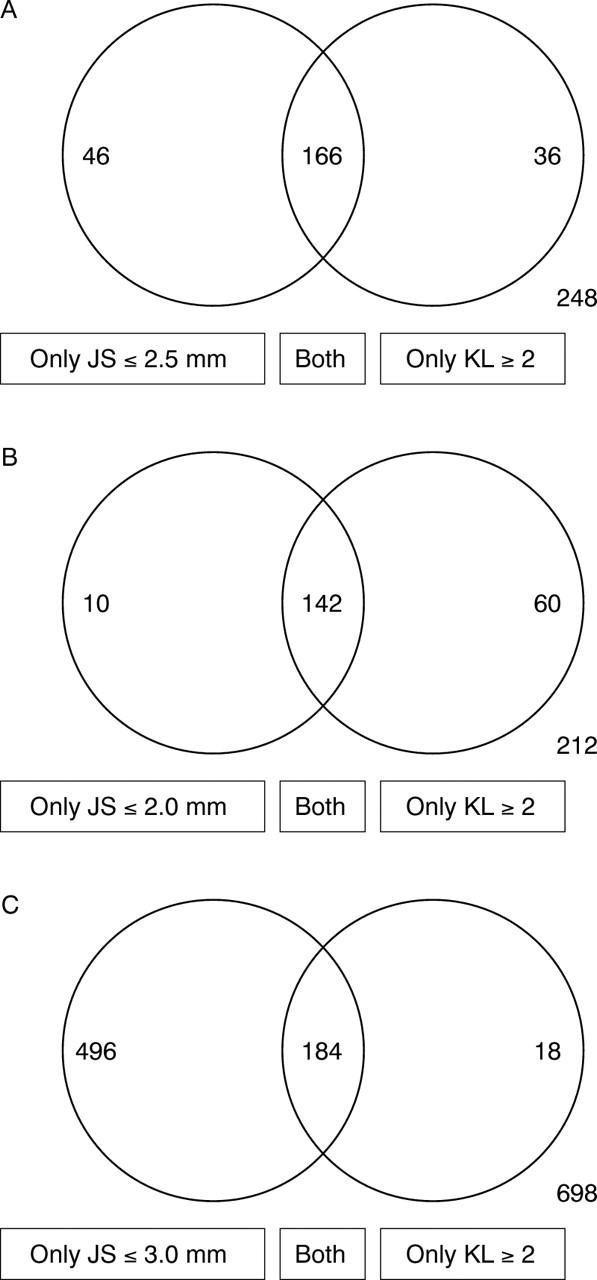
OBJECTIVE—To compare the reliability of quantitative measurement of minimum hip joint space with a qualitative global assessment of radiological features for estimating the prevalence of primary osteoarthritis (OA) of the hip in colon radiographs. METHODS—All colon radiographs from patients aged 35 or older, taken at three different radiographic departments in Iceland during the years 1990-96, were examined. A total of 3002 hips in 638 men and 863 women were analysed. Intraobserver and interobserver reliability was assessed by measuring 147 randomly selected radiographs (294 hips) twice by the same observer, and 87 and 98 randomly selected radiographs (174 and 196 hips) by two additional independent observers. Minimum hip joint space was measured with a millimetre ruler, and global assessment of radiological features by a published atlas. RESULTS—With a minimum joint space of 2.5 mm or less as definition for OA, 212 hips were defined as having OA. When the global Kellgren and Lawrence assessment with grade 2 (definite narrowing in the presence of definite osteophytes) or higher as definition for OA was used, 202 hips showed OA. However, only 166 hips were diagnosed as OA with both systems. With 2.0 or 3.0 mm minimum joint space as cut off point, the difference between the two methods increased. Both intrarater and interrater reliability was significantly higher with joint space measurement than with global assessment. CONCLUSIONS—Overall prevalence of radiological OA was similar with the two methods. However, the quantitative measurement of minimum hip joint space had a better within-observer and between-observer reliability than qualitative global assessment of radiographic features of hip OA. It is thus suggested that minimum joint space measurement is a preferable method in epidemiological studies of radiological hip OA.
Full Text
The Full Text of this article is available as a PDF (129.3 KB).
Figure 1 .

Agreement between the two methods for assessment of radiological hip OA. (A) Definition for radiological hip OA: joint space (JS) ⩽2.5 mm. (B) Definition for radiological hip OA: joint space ⩽2.0 mm. (C) Definition for radiological hip OA: joint space ⩽3.0 mm. Left circle: number of hips graded as having OA only with quantitative measurement of joint space. Right circle: number of hips graded as having OA only with qualitative assessment of radiological features by atlas (definite joint space narrowing in the presence of definite osteophytes). Overlapping circles: number of hips graded as having OA by both methods. Numbers outside circles: sum of number of hips assessed as having OA by any of the two methods.


