Abstract
OBJECTIVES—To (a) develop an atlas of line drawings for the assessment and grading of narrowing and osteophyte (that is, changes of osteoarthritis) on knee radiographs, and (b) compare the performance of this atlas with that of the standard Osteoarthritis Research Society (OARS) photographic atlas of radiographs. METHODS—Normal joint space widths (grade 0) for the medial and lateral tibiofemoral and medial and lateral patellofemoral compartments were obtained from a previous community study. Grades 1-3 narrowing in each compartment was calculated separately for men and women, grade 3 being bone on bone, grades 1 and 2 being two thirds and one third the value of grade 0. Maximum osteophyte size (grade 3) for each of eight sites was determined from 715 bilateral knee x ray films obtained in a knee osteoarthritis (OA) hospital clinic; grades 1-2 were calculated as two thirds and one third reductions in the area of grade 3. Drawings for narrowing and osteophyte were presented separately. 50 sets of bilateral knee x ray radiographs (standing, extended anteroposterior; flexed skyline) showing a spectrum of OA grades were scored by three observers, twice using the OARS atlas and twice using the drawn atlas. RESULTS—Intraobserver and interobserver reproducibility was similar and generally good with both atlases, though varied according to site. All three observers preferred the line drawing atlas for ease and convenience of use. Higher scores for patellofemoral narrowing and lower scores for osteophyte, especially medial femoral osteophyte, were seen using the line drawing atlas, showing that the two atlases are not equivalent instruments. CONCLUSION—A logically derived line drawing atlas for grading of narrowing and osteophyte at the knee has been produced. The atlas showed comparable reproducibility with the OARS atlas, but was discordant in several aspects of grading. Such a system has several theoretical and practical advantages and should be considered for use in knee OA studies.
Full Text
The Full Text of this article is available as a PDF (195.0 KB).
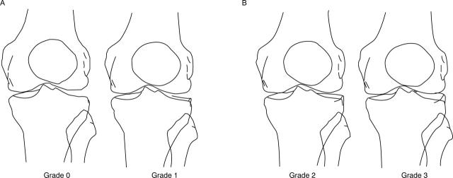
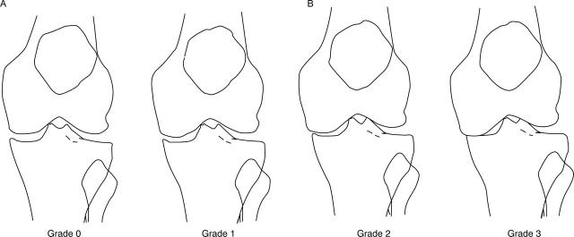
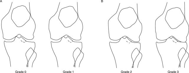
Figure 1 .
Medial tibiofemoral joint space narrowing for women. (A) Grades 0 and 1; (B) grades 2 and 3.
Figure 2 .
Lateral tibiofemoral joint space narrowing for women. (A) Grades 0 and 1; (B) grades 2 and 3.
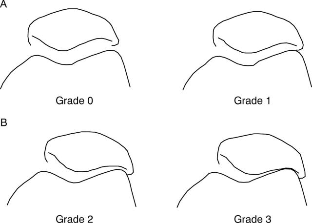
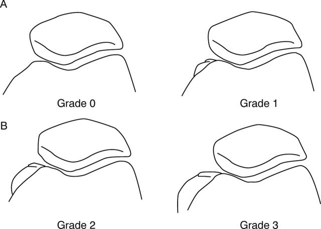
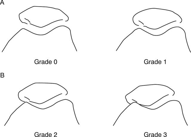
Figure 3 .
Lateral patellofemoral joint space narrowing for women. (A) Grades 0 and 1; (B) grades 2 and 3. Figure 4 Medial patellofemoral joint space narrowing for women. (A) Grades 0 and 1; (B) grades 2 and 3.
Figure 5 .
Osteophytes in all tibiofemoral sites. (A) grades 0 and 1; (B) grades 2 and 3.
Figure 6 .
Lateral tibial osteophyte (optional shape). These figures are applicable when the shape of lateral tibial osteophyte is considerably different from that of fig 5. (A) Grades 0 and 1; (B) grades 2 and 3.
Figure 7 .
Osteophyte in all patellofemoral sites. (A) Grades 0 and 1; (B) grades 2 and 3. Figure 8 Medial femoral trochlear osteophyte (optional shape). These figures are applicable when the shape of the medial femoral trochlea osteophyte is considerably different from that of fig 7. (A) Grades 0 and 1; (B) grades 2 and 3.
Figure 9 .
Medial tibiofemoral joint space narrowing for men. (A) Grades 0 and 1; (B) grades 2 and 3.
Figure 10 .
Lateral tibiofemoral joint space narrowing for men. (A) Grades 0 and 1; (B) grades 2 and 3.
Figure 11 .
Lateral patellofemoral joint space narrowing for men. (A) Grades 0 and 1; (B) grades 2 and 3. Figure 12 Medial patellofemoral joint space narrowing for men. (A) Grades 0 and 1; (B) grades 2 and 3.
Selected References
These references are in PubMed. This may not be the complete list of references from this article.
- Altman R. D., Fries J. F., Bloch D. A., Carstens J., Cooke T. D., Genant H., Gofton P., Groth H., McShane D. J., Murphy W. A. Radiographic assessment of progression in osteoarthritis. Arthritis Rheum. 1987 Nov;30(11):1214–1225. doi: 10.1002/art.1780301103. [DOI] [PubMed] [Google Scholar]
- Altman R. D., Hochberg M., Murphy W. A., Jr, Wolfe F., Lequesne M. Atlas of individual radiographic features in osteoarthritis. Osteoarthritis Cartilage. 1995 Sep;3 (Suppl A):3–70. [PubMed] [Google Scholar]
- Boegård T., Rudling O., Petersson I. F., Jonsson K. Correlation between radiographically diagnosed osteophytes and magnetic resonance detected cartilage defects in the patellofemoral joint. Ann Rheum Dis. 1998 Jul;57(7):395–400. doi: 10.1136/ard.57.7.395. [DOI] [PMC free article] [PubMed] [Google Scholar]
- Boegård T., Rudling O., Petersson I. F., Jonsson K. Correlation between radiographically diagnosed osteophytes and magnetic resonance detected cartilage defects in the tibiofemoral joint. Ann Rheum Dis. 1998 Jul;57(7):401–407. doi: 10.1136/ard.57.7.401. [DOI] [PMC free article] [PubMed] [Google Scholar]
- Brennan P., Silman A. Statistical methods for assessing observer variability in clinical measures. BMJ. 1992 Jun 6;304(6840):1491–1494. doi: 10.1136/bmj.304.6840.1491. [DOI] [PMC free article] [PubMed] [Google Scholar]
- Cicuttini F. M., Baker J., Hart D. J., Spector T. D. Choosing the best method for radiological assessment of patellofemoral osteoarthritis. Ann Rheum Dis. 1996 Feb;55(2):134–136. doi: 10.1136/ard.55.2.134. [DOI] [PMC free article] [PubMed] [Google Scholar]
- Cooper C., Cushnaghan J., Kirwan J. R., Dieppe P. A., Rogers J., McAlindon T., McCrae F. Radiographic assessment of the knee joint in osteoarthritis. Ann Rheum Dis. 1992 Jan;51(1):80–82. doi: 10.1136/ard.51.1.80. [DOI] [PMC free article] [PubMed] [Google Scholar]
- Dieppe P. A. Recommended methodology for assessing the progression of osteoarthritis of the hip and knee joints. Osteoarthritis Cartilage. 1995 Jun;3(2):73–77. doi: 10.1016/s1063-4584(05)80040-8. [DOI] [PubMed] [Google Scholar]
- Doherty M., Lanyon P. Epidemiology of peripheral joint osteoarthritis. Ann Rheum Dis. 1996 Sep;55(9):585–587. doi: 10.1136/ard.55.9.585. [DOI] [PMC free article] [PubMed] [Google Scholar]
- Dougados M., Gueguen A., Nguyen M., Thiesce A., Listrat V., Jacob L., Nakache J. P., Gabriel K. R., Lequesne M., Amor B. Longitudinal radiologic evaluation of osteoarthritis of the knee. J Rheumatol. 1992 Mar;19(3):378–384. [PubMed] [Google Scholar]
- Felson D. T. Epidemiology of hip and knee osteoarthritis. Epidemiol Rev. 1988;10:1–28. doi: 10.1093/oxfordjournals.epirev.a036019. [DOI] [PubMed] [Google Scholar]
- Günther K. P., Sun Y. Reliability of radiographic assessment in hip and knee osteoarthritis. Osteoarthritis Cartilage. 1999 Mar;7(2):239–246. doi: 10.1053/joca.1998.0152. [DOI] [PubMed] [Google Scholar]
- Jones A. C., Ledingham J., McAlindon T., Regan M., Hart D., MacMillan P. J., Doherty M. Radiographic assessment of patellofemoral osteoarthritis. Ann Rheum Dis. 1993 Sep;52(9):655–658. doi: 10.1136/ard.52.9.655. [DOI] [PMC free article] [PubMed] [Google Scholar]
- KELLGREN J. H., LAWRENCE J. S. Radiological assessment of osteo-arthrosis. Ann Rheum Dis. 1957 Dec;16(4):494–502. doi: 10.1136/ard.16.4.494. [DOI] [PMC free article] [PubMed] [Google Scholar]
- Kallman D. A., Wigley F. M., Scott W. W., Jr, Hochberg M. C., Tobin J. D. New radiographic grading scales for osteoarthritis of the hand. Reliability for determining prevalence and progression. Arthritis Rheum. 1989 Dec;32(12):1584–1591. doi: 10.1002/anr.1780321213. [DOI] [PubMed] [Google Scholar]
- Lanyon P., Jones A., Doherty M. Assessing progression of patellofemoral osteoarthritis: a comparison between two radiographic methods. Ann Rheum Dis. 1996 Dec;55(12):875–879. doi: 10.1136/ard.55.12.875. [DOI] [PMC free article] [PubMed] [Google Scholar]
- Lanyon P., O'Reilly S., Jones A., Doherty M. Radiographic assessment of symptomatic knee osteoarthritis in the community: definitions and normal joint space. Ann Rheum Dis. 1998 Oct;57(10):595–601. doi: 10.1136/ard.57.10.595. [DOI] [PMC free article] [PubMed] [Google Scholar]
- Laurin C. A., Dussault R., Levesque H. P. The tangential x-ray investigation of the patellofemoral joint: x-ray technique, diagnostic criteria and their interpretation. Clin Orthop Relat Res. 1979 Oct;(144):16–26. [PubMed] [Google Scholar]
- Ledingham J., Regan M., Jones A., Doherty M. Factors affecting radiographic progression of knee osteoarthritis. Ann Rheum Dis. 1995 Jan;54(1):53–58. doi: 10.1136/ard.54.1.53. [DOI] [PMC free article] [PubMed] [Google Scholar]
- McAlindon T. E., Cooper C., Kirwan J. R., Dieppe P. A. Knee pain and disability in the community. Br J Rheumatol. 1992 Mar;31(3):189–192. doi: 10.1093/rheumatology/31.3.189. [DOI] [PubMed] [Google Scholar]
- McAlindon T. E., Snow S., Cooper C., Dieppe P. A. Radiographic patterns of osteoarthritis of the knee joint in the community: the importance of the patellofemoral joint. Ann Rheum Dis. 1992 Jul;51(7):844–849. doi: 10.1136/ard.51.7.844. [DOI] [PMC free article] [PubMed] [Google Scholar]
- O'Reilly S. C., Jones A., Muir K. R., Doherty M. Quadriceps weakness in knee osteoarthritis: the effect on pain and disability. Ann Rheum Dis. 1998 Oct;57(10):588–594. doi: 10.1136/ard.57.10.588. [DOI] [PMC free article] [PubMed] [Google Scholar]
- Schouten J. S., van den Ouweland F. A., Valkenburg H. A. A 12 year follow up study in the general population on prognostic factors of cartilage loss in osteoarthritis of the knee. Ann Rheum Dis. 1992 Aug;51(8):932–937. doi: 10.1136/ard.51.8.932. [DOI] [PMC free article] [PubMed] [Google Scholar]
- Scott W. W., Jr, Lethbridge-Cejku M., Reichle R., Wigley F. M., Tobin J. D., Hochberg M. C. Reliability of grading scales for individual radiographic features of osteoarthritis of the knee. The Baltimore longitudinal study of aging atlas of knee osteoarthritis. Invest Radiol. 1993 Jun;28(6):497–501. [PubMed] [Google Scholar]
- Spector T. D., Cooper C. Radiographic assessment of osteoarthritis in population studies: whither Kellgren and Lawrence? Osteoarthritis Cartilage. 1993 Oct;1(4):203–206. doi: 10.1016/s1063-4584(05)80325-5. [DOI] [PubMed] [Google Scholar]
- Spector T. D., Hart D. J., Byrne J., Harris P. A., Dacre J. E., Doyle D. V. Definition of osteoarthritis of the knee for epidemiological studies. Ann Rheum Dis. 1993 Nov;52(11):790–794. doi: 10.1136/ard.52.11.790. [DOI] [PMC free article] [PubMed] [Google Scholar]
- Sun Y., Günther K. P., Brenner H. Reliability of radiographic grading of osteoarthritis of the hip and knee. Scand J Rheumatol. 1997;26(3):155–165. doi: 10.3109/03009749709065675. [DOI] [PubMed] [Google Scholar]


