

Abstract
OBJECTIVE—To determine the levels of vascular endothelial growth factor (VEGF) mRNA and protein expression in normal and osteoarthritic (OA) human articular cartilage, and whether VEGF expression alters during the progression of OA. METHODS—Sections from normal and OA human knee cartilage were immunotained with a polyclonal antibody recognising VEGF. In addition, total RNA was isolated from normal and osteoarthritic human knee cartilage and analysed by reverse transcriptase-polymerase chain reaction (RT-PCR) for VEGF mRNA expression. RESULTS—VEGF was found to be present in normal and OA human knee cartilage in all cartilage layers. A significant increase of VEGF immunopositive chondrocytes to up to ~82% was detected in severe OA cartilage compared with normal articular cartilage (~56% of immunopositive chondrocytes). RT-PCR analysis showed the expression of VEGF also on the mRNA level. CONCLUSIONS—VEGF is expressed by articular chondrocytes in normal and OA human knee cartilage. The percentage of VEGF immunopositive chondrocytes significantly increases in late stages of the disease. The VEGF transcript levels encoding all four isoforms shows a big variability in samples from different donors, suggesting a distinct regulation of the expression of the four VEGF isoforms in normal and OA cartilage.
Full Text
The Full Text of this article is available as a PDF (151.1 KB).
Figure 1 .
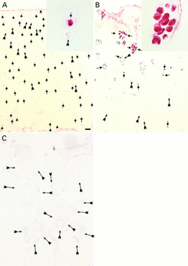
(A) Normal articular cartilage immunostained with antibodies against VEGF, showing positive (arrowheads) and negative chondrocytes (arrows) in all cartilage layers; Mankin score 1. VEGF positive and negative chondrocytes from the deep zone are shown in close up (inset). (B) Severe OA cartilage with a strong immunostaining of the chondrocyte clusters next to the surface (arrowheads) and also VEGF positive chondrocytes throughout the remaining deep zone (arrows); Mankin score 11. Positive chondrocytes within a cluster are displayed in close up (inset). (C) A slide adjacent to (B) incubated with non-immune rabbit serum as negative control. Arrows indicate negative chondrocytes. Bar represents: (A) 200 µm; (B) 100 µm; and (C) 60 µm.
Figure 2 .

Reverse transcriptase-polymerase chain reaction (RT-PCR) analyses of VEGF mRNA levels compared with corresponding GAPDH mRNA levels in human normal and OA samples. Resulting PCR products were analysed on a 1.5% agarose gel. (A) VEGF in normal cartilage. Lanes 1 and 2 represent the PCR product (378 bp) of the same sample with 35 or 29 cycles, respectively. (B) PCR products of different OA cartilage samples after 35 cycles; lane 1 represents a grade I lesion, lanes 2 and 3 show grade II lesions, lanes 5 and 6 demonstrate grade III lesions. (C and D) represent the corresponding GAPDH PCR products (294 bp) of the same donors as shown above.