Abstract
OBJECTIVE—To evaluate the efficacy of extracorporeal shock wave treatment (ESWT) in calcaneal enthesophytosis. METHODS—60 patients (43 women, 17 men) were examined who had talalgia associated with heel spur. A single blind randomised study was performed in which 30 patients underwent a regular treatment (group 1) and 30 a simulated one (shocks of 0 mJ/mm2 energy were applied) (group 2). Variations in symptoms were evaluated by visual analogue scale (VAS). Variations in the dimension of enthesophytosis were evaluated by x ray examination. Variations in the grade of enthesitis were evaluated by sonography. RESULTS—A significant decrease of VAS was seen in group 1. Examination by x ray showed morphological modifications (reduction of the larger diameter >1 mm) of the enthesophytosis in nine (30%) patients. Sonography did not show significant changes in the grade of enthesitis just after the end of the treatment, but a significant reduction was seen after one month. In the control group no significant decrease of VAS was seen. No modification was observed by x ray examination or sonography. CONCLUSION—ESWT is safe and improves the symptoms of most patients with a painful heel, it can also structurally modify enthesophytosis, and reduce inflammatory oedema.
Full Text
The Full Text of this article is available as a PDF (153.1 KB).
Figure 1 .
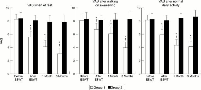
Mean visual analogue scale (VAS) score before extracorporeal shock wave treatment (ESWT), after ESWT, and one and three months later, at rest, after walking on awakening, and after normal daily activity. *p<0.0001, group 1 v group 2; †p<0.0001 group 1, baseline v after ESWT, after one month, and after three months.
Figure 2 .

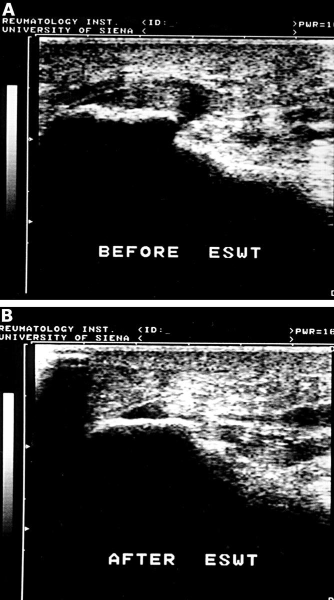
(A) Enthesitis of the plantar fascia (sonographic grade 3) before extracorporeal shock wave treatment (ESWT). (B) The same patient as in fig 2A, examined one month after the end of ESWT. An improvement of enthesitis (disappearance of the paratendinitis, reduction of thickness of the plantar fascia origin, modification of the subcalcaneal spur) can be seen.
Figure 3 .
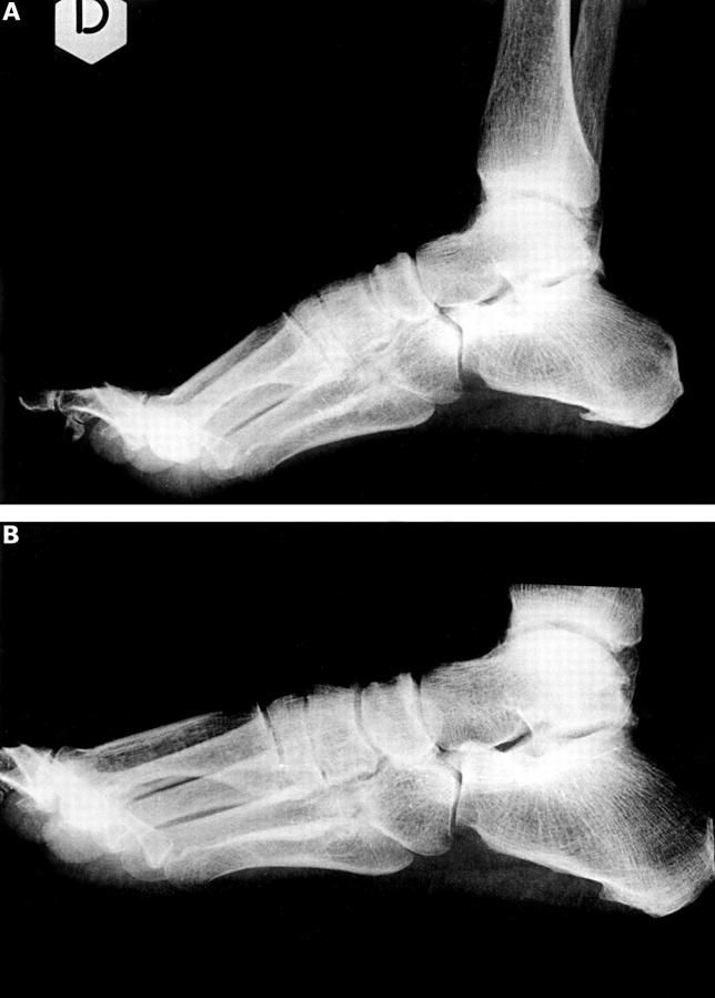
(A) An x ray picture of heel spur before extracorporeal shock wave treatment. (B) The same patient as in fig 3A one month after the end of the treatment.


