

Abstract
BACKGROUND—Volumes of inflamed synovial membrane determined by magnetic resonance imaging (MRI) are closely related to histopathological synovitis and may predict erosive progression in rheumatoid arthritis (RA). However, after IV injection, leakage of MRI contrast from the synovium gradually compromises the differentiation of synovium from joint fluid. OBJECTIVE—To determine the time period after IV MRI contrast (gadolinium-DTPA (Gd)) injection in which synovial membrane volume determination is reliable. METHODS—MRI of five RA knees with clinical synovitis was carried out, with axial, T1 weighted, spin echo images before IV Gd injection and every 1.75 minutes for 60 minutes post-Gd. By a semiautomated "signal enhancement threshold" method, including voxels with >35% or >45% relative post-Gd enhancement, synovial membrane volumes were estimated at each time point. At 4.25 minutes post-Gd, volumes were also determined by a more accurate but time consuming "manual method". RESULTS—The initially observed synovium-effusion borderline remained clearly visible, and on the same location, within at least the initial 11 minutes post-Gd (that is, within the normal time frame of post-Gd imaging in RA) but started blurring and moving centripetally thereafter. Compared with volumes at all other time points, synovial membrane volumes at 0.75 and 2.50 minutes post-Gd were significantly lower (Wilcoxon-Pratt), suggesting that some synovial membrane areas had not yet exceeded the enhancement threshold. Thereafter, the measured volumes remained practically unchanged. CONCLUSION—This study suggests that MR image acquisition in arthritic knee joints should be performed within the initial approximately 10 minutes after gadolinium contrast injection to achieve the most accurate distinction between synovium and joint fluid but that small time variations are not of major importance to the measured synovial membrane volumes.
Full Text
The Full Text of this article is available as a PDF (225.1 KB).
Figure 1 .
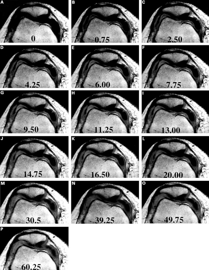
Axial T1 weighted MR images through the patella and the parapatellar recesses, (A) before and (B-P) at successive time points after IV gadolinium-DTPA. Numbers indicate the time (minutes) after contrast injection. A signal intensity increase (enhancement) is seen immediately in the synovial membrane, while the joint fluid enhances gradually, from the periphery.
Figure 2 .
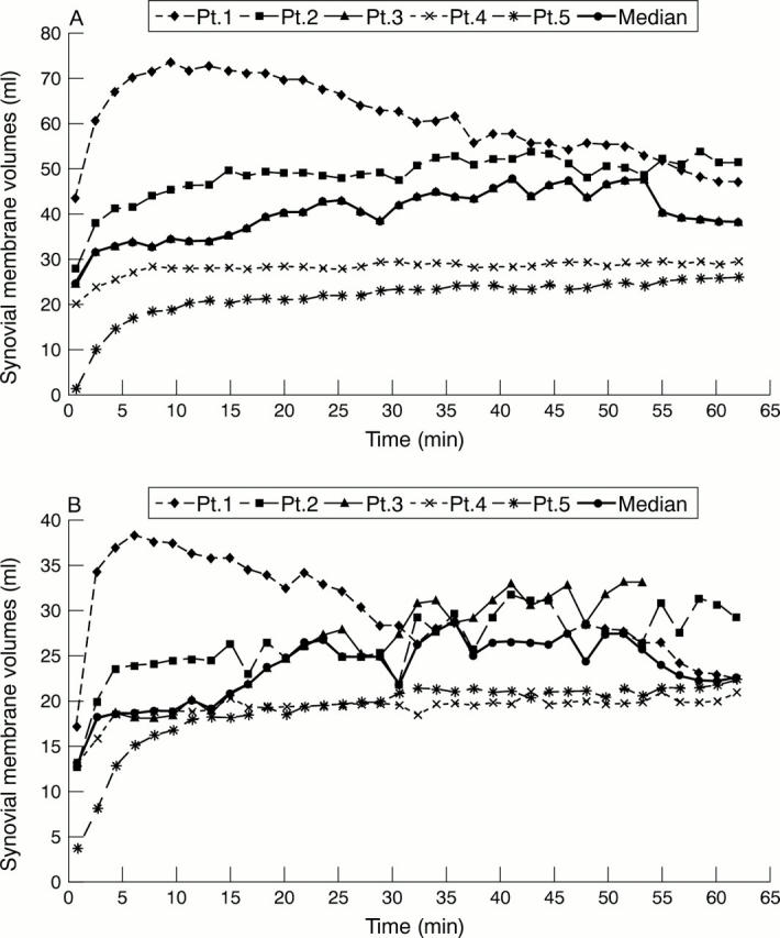
The course of the synovial membrane volume, as estimated by the enhancement threshold method, within the first 60 minutes after IV gadolinium-DTPA. (A) Enhancement threshold >35%; (B) enhancement threshold >45%.
Selected References
These references are in PubMed. This may not be the complete list of references from this article.
- Arnett F. C., Edworthy S. M., Bloch D. A., McShane D. J., Fries J. F., Cooper N. S., Healey L. A., Kaplan S. R., Liang M. H., Luthra H. S. The American Rheumatism Association 1987 revised criteria for the classification of rheumatoid arthritis. Arthritis Rheum. 1988 Mar;31(3):315–324. doi: 10.1002/art.1780310302. [DOI] [PubMed] [Google Scholar]
- Clunie G., Hall-Craggs M. A., Paley M. N., King A., Wilkinson I. D., Ell P. J., Edwards J. C. Measurement of synovial lining volume by magnetic resonance imaging of the knee in chronic synovitis. Ann Rheum Dis. 1997 Sep;56(9):526–534. doi: 10.1136/ard.56.9.526. [DOI] [PMC free article] [PubMed] [Google Scholar]
- Creamer P., Keen M., Zananiri F., Waterton J. C., Maciewicz R. A., Oliver C., Dieppe P., Watt I. Quantitative magnetic resonance imaging of the knee: a method of measuring response to intra-articular treatments. Ann Rheum Dis. 1997 Jun;56(6):378–381. doi: 10.1136/ard.56.6.378. [DOI] [PMC free article] [PubMed] [Google Scholar]
- Drapé J. L., Thelen P., Gay-Depassier P., Silbermann O., Benacerraf R. Intraarticular diffusion of Gd-DOTA after intravenous injection in the knee: MR imaging evaluation. Radiology. 1993 Jul;188(1):227–234. doi: 10.1148/radiology.188.1.8511303. [DOI] [PubMed] [Google Scholar]
- Gaffney K., Cookson J., Blake D., Coumbe A., Blades S. Quantification of rheumatoid synovitis by magnetic resonance imaging. Arthritis Rheum. 1995 Nov;38(11):1610–1617. doi: 10.1002/art.1780381113. [DOI] [PubMed] [Google Scholar]
- Hervé-Somma C. M., Sebag G. H., Prieur A. M., Bonnerot V., Lallemand D. P. Juvenile rheumatoid arthritis of the knee: MR evaluation with Gd-DOTA. Radiology. 1992 Jan;182(1):93–98. doi: 10.1148/radiology.182.1.1727317. [DOI] [PubMed] [Google Scholar]
- Kursunoglu-Brahme S., Riccio T., Weisman M. H., Resnick D., Zvaifler N., Sanders M. E., Fix C. Rheumatoid knee: role of gadopentetate-enhanced MR imaging. Radiology. 1990 Sep;176(3):831–835. doi: 10.1148/radiology.176.3.2389044. [DOI] [PubMed] [Google Scholar]
- König H., Sieper J., Wolf K. J. Rheumatoid arthritis: evaluation of hypervascular and fibrous pannus with dynamic MR imaging enhanced with Gd-DTPA. Radiology. 1990 Aug;176(2):473–477. doi: 10.1148/radiology.176.2.2367663. [DOI] [PubMed] [Google Scholar]
- Ostergaard M., Court-Payen M., Gideon P., Wieslander S., Cortsen M., Lorenzen I., Henriksen O. Ultrasonography in arthritis of the knee. A comparison with MR imaging. Acta Radiol. 1995 Jan;36(1):19–26. [PubMed] [Google Scholar]
- Ostergaard M. Different approaches to synovial membrane volume determination by magnetic resonance imaging: manual versus automated segmentation. Br J Rheumatol. 1997 Nov;36(11):1166–1177. doi: 10.1093/rheumatology/36.11.1166. [DOI] [PubMed] [Google Scholar]
- Ostergaard M., Ejbjerg B., Stoltenberg M., Gideon P., Volck B., Skov K., Jensen C. H., Lorenzen I. Quantitative magnetic resonance imaging as marker of synovial membrane regeneration and recurrence of synovitis after arthroscopic knee joint synovectomy: a one year follow up study. Ann Rheum Dis. 2001 Mar;60(3):233–236. doi: 10.1136/ard.60.3.233. [DOI] [PMC free article] [PubMed] [Google Scholar]
- Ostergaard M., Gideon P., Henriksen O., Lorenzen I. Synovial volume--a marker of disease severity in rheumatoid arthritis? Quantification by MRI. Scand J Rheumatol. 1994;23(4):197–202. doi: 10.3109/03009749409103060. [DOI] [PubMed] [Google Scholar]
- Ostergaard M., Hansen M., Stoltenberg M., Gideon P., Klarlund M., Jensen K. E., Lorenzen I. Magnetic resonance imaging-determined synovial membrane volume as a marker of disease activity and a predictor of progressive joint destruction in the wrists of patients with rheumatoid arthritis. Arthritis Rheum. 1999 May;42(5):918–929. doi: 10.1002/1529-0131(199905)42:5<918::AID-ANR10>3.0.CO;2-2. [DOI] [PubMed] [Google Scholar]
- Ostergaard M. Magnetic resonance imaging in rheumatoid arthritis. Quantitative methods for assessment of the inflammatory process in peripheral joints. Dan Med Bull. 1999 Sep;46(4):313–344. [PubMed] [Google Scholar]
- Ostergaard M., Stoltenberg M., Gideon P., Sørensen K., Henriksen O., Lorenzen I. Changes in synovial membrane and joint effusion volumes after intraarticular methylprednisolone. Quantitative assessment of inflammatory and destructive changes in arthritis by MRI. J Rheumatol. 1996 Jul;23(7):1151–1161. [PubMed] [Google Scholar]
- Ostergaard M., Stoltenberg M., Henriksen O., Lorenzen I. Quantitative assessment of synovial inflammation by dynamic gadolinium-enhanced magnetic resonance imaging. A study of the effect of intra-articular methylprednisolone on the rate of early synovial enhancement. Br J Rheumatol. 1996 Jan;35(1):50–59. doi: 10.1093/rheumatology/35.1.50. [DOI] [PubMed] [Google Scholar]
- Ostergaard M., Stoltenberg M., Henriksen O., Lorenzen I. The accuracy of MRI-determined synovial membrane and joint effusion volumes in arthritis. A comparison of pre- and post-aspiration volumes. Scand J Rheumatol. 1995;24(5):305–311. doi: 10.3109/03009749509095168. [DOI] [PubMed] [Google Scholar]
- Ostergaard M., Stoltenberg M., Løvgreen-Nielsen P., Volck B., Jensen C. H., Lorenzen I. Magnetic resonance imaging-determined synovial membrane and joint effusion volumes in rheumatoid arthritis and osteoarthritis: comparison with the macroscopic and microscopic appearance of the synovium. Arthritis Rheum. 1997 Oct;40(10):1856–1867. doi: 10.1002/art.1780401020. [DOI] [PubMed] [Google Scholar]
- Palmer W. E., Rosenthal D. I., Schoenberg O. I., Fischman A. J., Simon L. S., Rubin R. H., Polisson R. P. Quantification of inflammation in the wrist with gadolinium-enhanced MR imaging and PET with 2-[F-18]-fluoro-2-deoxy-D-glucose. Radiology. 1995 Sep;196(3):647–655. doi: 10.1148/radiology.196.3.7644624. [DOI] [PubMed] [Google Scholar]
- Peterfy C. G., Majumdar S., Lang P., van Dijke C. F., Sack K., Genant H. K. MR imaging of the arthritic knee: improved discrimination of cartilage, synovium, and effusion with pulsed saturation transfer and fat-suppressed T1-weighted sequences. Radiology. 1994 May;191(2):413–419. doi: 10.1148/radiology.191.2.8153315. [DOI] [PubMed] [Google Scholar]
- Polisson R. P., Schoenberg O. I., Fischman A., Rubin R., Simon L. S., Rosenthal D., Palmer W. E. Use of magnetic resonance imaging and positron emission tomography in the assessment of synovial volume and glucose metabolism in patients with rheumatoid arthritis. Arthritis Rheum. 1995 Jun;38(6):819–825. doi: 10.1002/art.1780380616. [DOI] [PubMed] [Google Scholar]
- Reiser M. F., Bongartz G. P., Erlemann R., Schneider M., Pauly T., Sittek H., Peters P. E. Gadolinium-DTPA in rheumatoid arthritis and related diseases: first results with dynamic magnetic resonance imaging. Skeletal Radiol. 1989;18(8):591–597. doi: 10.1007/BF00355334. [DOI] [PubMed] [Google Scholar]
- Sugimoto H., Takeda A., Kano S. Assessment of disease activity in rheumatoid arthritis using magnetic resonance imaging: quantification of pannus volume in the hands. Br J Rheumatol. 1998 Aug;37(8):854–861. doi: 10.1093/rheumatology/37.8.854. [DOI] [PubMed] [Google Scholar]
- Waterton J. C., Rajanayagam V., Ross B. D., Brown D., Whittemore A., Johnstone D. Magnetic resonance methods for measurement of disease progression in rheumatoid arthritis. Magn Reson Imaging. 1993;11(7):1033–1038. doi: 10.1016/0730-725x(93)90222-y. [DOI] [PubMed] [Google Scholar]
- Winalski C. S., Aliabadi P., Wright R. J., Shortkroff S., Sledge C. B., Weissman B. N. Enhancement of joint fluid with intravenously administered gadopentetate dimeglumine: technique, rationale, and implications. Radiology. 1993 Apr;187(1):179–185. doi: 10.1148/radiology.187.1.8451409. [DOI] [PubMed] [Google Scholar]
- Winalski C. S., Palmer W. E., Rosenthal D. I., Weissman B. N. Magnetic resonance imaging of rheumatoid arthritis. Radiol Clin North Am. 1996 Mar;34(2):243-58, x. [PubMed] [Google Scholar]
- Yamato M., Tamai K., Yamaguchi T., Ohno W. MRI of the knee in rheumatoid arthritis: Gd-DTPA perfusion dynamics. J Comput Assist Tomogr. 1993 Sep-Oct;17(5):781–785. doi: 10.1097/00004728-199309000-00022. [DOI] [PubMed] [Google Scholar]