

Abstract
OBJECTIVES—By repeated magnetic resonance imaging (MRI) to study synovial membrane regeneration and recurrence of synovitis after arthroscopic knee joint synovectomy in patients with rheumatoid arthritis (RA) and other (non-RA) causes of persistent knee joint synovitis. METHODS—Contrast enhanced MRI was performed in 15 knees (nine RA, six non-RA) before and one day, seven days, two months, and 12 months after arthroscopic synovectomy. Synovial membrane volumes, joint effusion volumes, and cartilage and bone destruction were assessed on each MRI set. Baseline microscopic and macroscopic assessments of synovitis and baseline and follow up standard clinical and biochemical examinations were available. RESULTS—Synovial membrane and joint fluid volumes were significantly reduced two and 12 months after synovectomy. However, MRI signs of recurrent synovitis were already present in most knees at two months. No significant differences between volumes in RA and non-RA knees were seen. Synovial membrane volumes at two months were significantly inversely correlated with the duration of clinical remission, for all knees considered together (Spearman's correlation rs=−0.67; p<0.05), for RA knees (rs=−0.76; p<0.05), and for non-RA knees (rs=−0.83; p<0.05). Baseline volumes were not significantly correlated with clinical outcome. Only three knees (all RA) showed erosive progression. The rate of erosive progression was not correlated with MRI volumes or with clinical or biochemical parameters. CONCLUSION—The synovial membrane had regenerated two months after arthroscopic knee joint synovectomy and despite significant volume reductions compared with baseline it often showed signs of recurrent synovitis. MRI seems to be valuable as a marker of inflammation, destruction and, perhaps, as a predictor of therapeutic outcome in arthritis.
Full Text
The Full Text of this article is available as a PDF (153.5 KB).
Figure 1 .
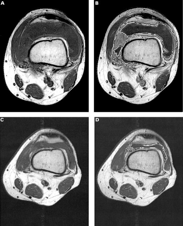
Transversal T1 weighted spin echo magnetic resonance images through the suprapatellar recess. (A, B) Before arthroscopic synovectomy, (A) before and (B) after intravenous injection of Gd-DTPA. After intravenous Gd-DTPA the signal intensity of the synovial membrane (outlined) has increased markedly, while the joint fluid remains dark grey. MRI determined synovial membrane and joint fluid volumes were 177 cm3 and 173 cm3, respectively. (C, D) Corresponding images two months after arthroscopic synovectomy, showing regeneration of the synovial membrane and, despite markedly decreased signs of joint inflammation, some synovitis and joint effusion (volumes 45 cm3 and 12 cm3, respectively).
Selected References
These references are in PubMed. This may not be the complete list of references from this article.
- Arnett F. C., Edworthy S. M., Bloch D. A., McShane D. J., Fries J. F., Cooper N. S., Healey L. A., Kaplan S. R., Liang M. H., Luthra H. S. The American Rheumatism Association 1987 revised criteria for the classification of rheumatoid arthritis. Arthritis Rheum. 1988 Mar;31(3):315–324. doi: 10.1002/art.1780310302. [DOI] [PubMed] [Google Scholar]
- Combe B., Krause E., Sany J. Treatment of chronic knee synovitis with arthroscopic synovectomy after failure of intraarticular injection of radionuclide. Arthritis Rheum. 1989 Jan;32(1):10–14. doi: 10.1002/anr.1780320103. [DOI] [PubMed] [Google Scholar]
- Fiocco U., Cozzi L., Rigon C., Chieco-Bianchi F., Baldovin M., Cassisi G. A., Gallo C., Doria A., Favaro M. A., Piccoli A. Arthroscopic synovectomy in rheumatoid and psoriatic knee joint synovitis: long-term outcome. Br J Rheumatol. 1996 May;35(5):463–470. doi: 10.1093/rheumatology/35.5.463. [DOI] [PubMed] [Google Scholar]
- Fiocco U., Cozzi L., Rubaltelli L., Rigon C., De Candia A., Tregnaghi A., Gallo C., Favaro M. A., Chieco-Bianchi F., Baldovin M. Long-term sonographic follow-up of rheumatoid and psoriatic proliferative knee joint synovitis. Br J Rheumatol. 1996 Feb;35(2):155–163. doi: 10.1093/rheumatology/35.2.155. [DOI] [PubMed] [Google Scholar]
- Hervé-Somma C. M., Sebag G. H., Prieur A. M., Bonnerot V., Lallemand D. P. Juvenile rheumatoid arthritis of the knee: MR evaluation with Gd-DOTA. Radiology. 1992 Jan;182(1):93–98. doi: 10.1148/radiology.182.1.1727317. [DOI] [PubMed] [Google Scholar]
- Matsui N., Taneda Y., Ohta H., Itoh T., Tsuboguchi S. Arthroscopic versus open synovectomy in the rheumatoid knee. Int Orthop. 1989;13(1):17–20. doi: 10.1007/BF00266717. [DOI] [PubMed] [Google Scholar]
- McQueen F. M., Stewart N., Crabbe J., Robinson E., Yeoman S., Tan P. L., McLean L. Magnetic resonance imaging of the wrist in early rheumatoid arthritis reveals progression of erosions despite clinical improvement. Ann Rheum Dis. 1999 Mar;58(3):156–163. doi: 10.1136/ard.58.3.156. [DOI] [PMC free article] [PubMed] [Google Scholar]
- Ogilvie-Harris D. J., Basinski A. Arthroscopic synovectomy of the knee for rheumatoid arthritis. Arthroscopy. 1991;7(1):91–97. doi: 10.1016/0749-8063(91)90085-c. [DOI] [PubMed] [Google Scholar]
- Ostergaard M., Gideon P., Henriksen O., Lorenzen I. Synovial volume--a marker of disease severity in rheumatoid arthritis? Quantification by MRI. Scand J Rheumatol. 1994;23(4):197–202. doi: 10.3109/03009749409103060. [DOI] [PubMed] [Google Scholar]
- Ostergaard M., Hansen M., Stoltenberg M., Gideon P., Klarlund M., Jensen K. E., Lorenzen I. Magnetic resonance imaging-determined synovial membrane volume as a marker of disease activity and a predictor of progressive joint destruction in the wrists of patients with rheumatoid arthritis. Arthritis Rheum. 1999 May;42(5):918–929. doi: 10.1002/1529-0131(199905)42:5<918::AID-ANR10>3.0.CO;2-2. [DOI] [PubMed] [Google Scholar]
- Ostergaard M., Stoltenberg M., Gideon P., Sørensen K., Henriksen O., Lorenzen I. Changes in synovial membrane and joint effusion volumes after intraarticular methylprednisolone. Quantitative assessment of inflammatory and destructive changes in arthritis by MRI. J Rheumatol. 1996 Jul;23(7):1151–1161. [PubMed] [Google Scholar]
- Ostergaard M., Stoltenberg M., Henriksen O., Lorenzen I. The accuracy of MRI-determined synovial membrane and joint effusion volumes in arthritis. A comparison of pre- and post-aspiration volumes. Scand J Rheumatol. 1995;24(5):305–311. doi: 10.3109/03009749509095168. [DOI] [PubMed] [Google Scholar]
- Ostergaard M., Stoltenberg M., Løvgreen-Nielsen P., Volck B., Jensen C. H., Lorenzen I. Magnetic resonance imaging-determined synovial membrane and joint effusion volumes in rheumatoid arthritis and osteoarthritis: comparison with the macroscopic and microscopic appearance of the synovium. Arthritis Rheum. 1997 Oct;40(10):1856–1867. doi: 10.1002/art.1780401020. [DOI] [PubMed] [Google Scholar]
- Pages M., Poey C., Lassoued S., Fournie B., Railhac J. J. MR imaging of the knee in rheumatoid arthritis and other rheumatic diseases. AJR Am J Roentgenol. 1991 Nov;157(5):1128–1128. doi: 10.2214/ajr.157.5.1927804. [DOI] [PubMed] [Google Scholar]
- Poleksic L., Zdravkovic D., Jablanovic D., Watt I., Bacic G. Magnetic resonance imaging of bone destruction in rheumatoid arthritis: comparison with radiography. Skeletal Radiol. 1993 Nov;22(8):577–580. doi: 10.1007/BF00197138. [DOI] [PubMed] [Google Scholar]
- Smiley P., Wasilewski S. A. Arthroscopic synovectomy. Arthroscopy. 1990;6(1):18–23. doi: 10.1016/0749-8063(90)90091-q. [DOI] [PubMed] [Google Scholar]
- Waterton J. C., Rajanayagam V., Ross B. D., Brown D., Whittemore A., Johnstone D. Magnetic resonance methods for measurement of disease progression in rheumatoid arthritis. Magn Reson Imaging. 1993;11(7):1033–1038. doi: 10.1016/0730-725x(93)90222-y. [DOI] [PubMed] [Google Scholar]
- Yulish B. S., Lieberman J. M., Newman A. J., Bryan P. J., Mulopulos G. P., Modic M. T. Juvenile rheumatoid arthritis: assessment with MR imaging. Radiology. 1987 Oct;165(1):149–152. doi: 10.1148/radiology.165.1.3628761. [DOI] [PubMed] [Google Scholar]