Full Text
The Full Text of this article is available as a PDF (293.4 KB).
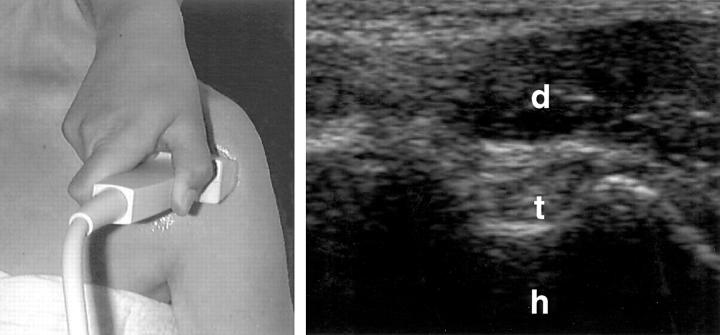
Figure 1 .
Anterior transverse scan in neutral position at the bicipital groove. h = humerus; t = biceps tendon; d = deltoid muscle.
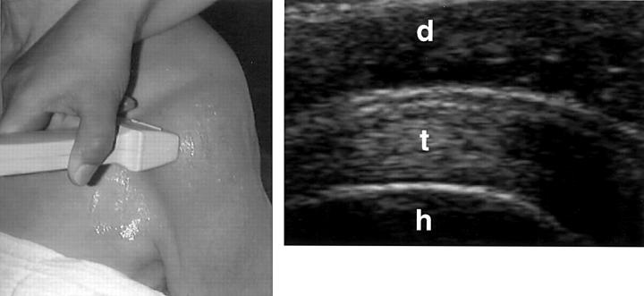
Figure 2 .
Anterior transverse scan in maximal internal rotation of the shoulder. h = humerus; t = supraspinatus tendon; d = deltoid muscle.
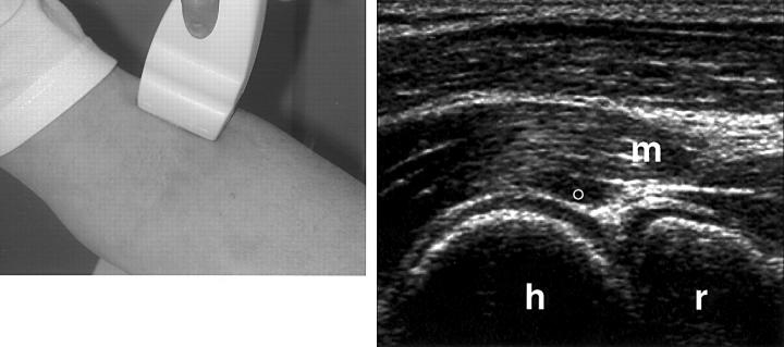
Figure 3 .
Anterior humeroradial longitudinal scan at the elbow. h = humerus; r = radius; m = muscles; ° = articular cartilage
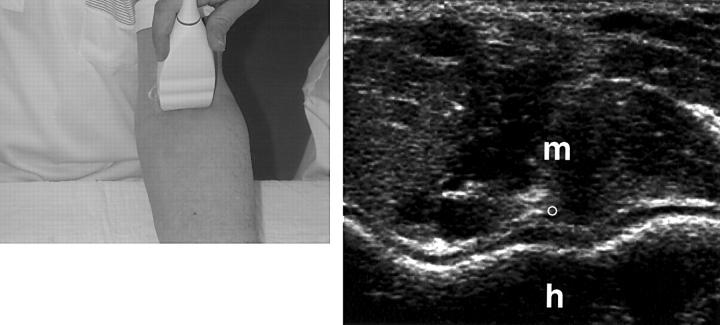
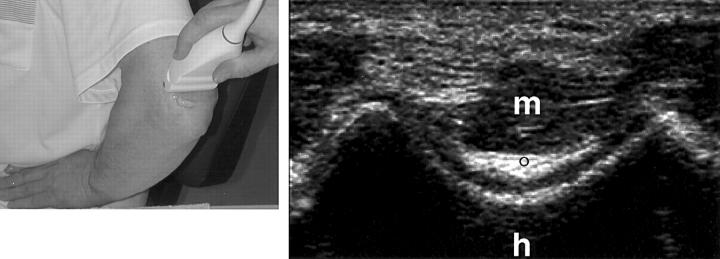
Figure 4 .
Anterior transverse scan at the distal humeral epiphysis. h = humerus; ° = articular cartilage; m = muscles.
Figure 5 .
Posterior transverse scan at the distal humeral epiphysis. h = humerus; ° = articular cartilage; m = triceps muscle.
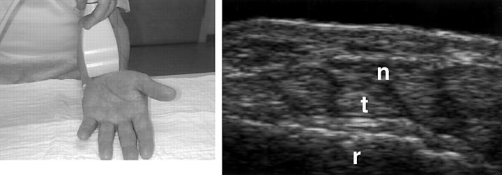
Figure 6 .
Volar transverse scan at the carpal tunnel. r = radius; n = median nerve; t = flexor tendons.
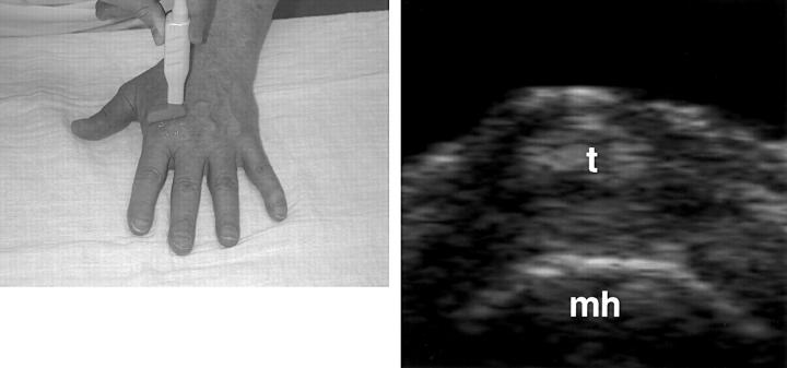
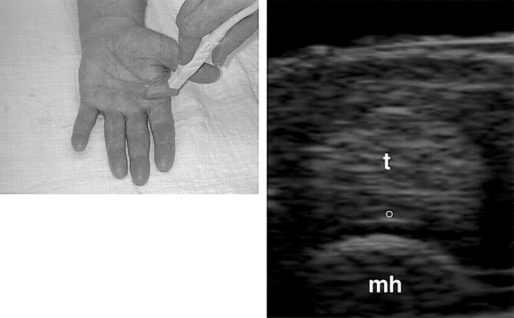
Figure 7 .
Dorsal transverse scan at the metacarpal head. mh = metacarpal head; t = extensor tendon.
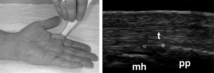
Figure 8 .
Palmar longitudinal scan at the metacarpophalangeal joint. * = joint cavity; ° = articular cartilage; pp = proximal phalanx; mh = metacarpal head; t = flexor tendon.
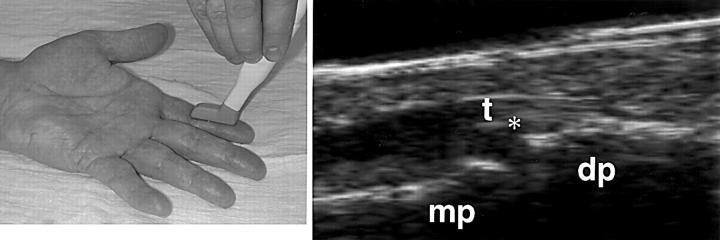
Figure 9 .
Palmar longitudinal scan at the distal interphalangeal joint. * = joint cavity; dp = proximal phalanx; mp = middle phalanx; t = flexor tendon.
Figure 10 .
Palmar transverse scan at the metacarpal head. mh = metacarpal head; ° = articular cartilage; t = flexor tendon.
Figure 11 .
Anterior longitudinal scan at the hip. a = acetabulum; f = femur; * = joint cavity; m = muscles.
Figure 12 .
Suprapatellar transverse scan in maximal flexion. f = femur; ° = articular cartilage.
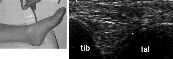
Figure 13 .
Anterior longitudinal scan at the ankle. tib = tibia; tal = talus; ° = articular cartilage.
Figure 14 .
Posterior longitudinal scan at the heel. t = achilles tendon; cal = calcaneus; k = Kager's fat pat.
Figure 15 .
Dorsal longitudinal scan at the first toe. mh = metatarsal head; ; pp = proximal phalanx; t = extensor tendon; * = joint cavity; ° = articular cartilage.
Selected References
These references are in PubMed. This may not be the complete list of references from this article.
- Backhaus M., Kamradt T., Sandrock D., Loreck D., Fritz J., Wolf K. J., Raber H., Hamm B., Burmester G. R., Bollow M. Arthritis of the finger joints: a comprehensive approach comparing conventional radiography, scintigraphy, ultrasound, and contrast-enhanced magnetic resonance imaging. Arthritis Rheum. 1999 Jun;42(6):1232–1245. doi: 10.1002/1529-0131(199906)42:6<1232::AID-ANR21>3.0.CO;2-3. [DOI] [PubMed] [Google Scholar]
- Gibbon W. W., Wakefield R. J. Ultrasound in inflammatory disease. Radiol Clin North Am. 1999 Jul;37(4):633–651. doi: 10.1016/s0033-8389(05)70120-2. [DOI] [PubMed] [Google Scholar]
- Grassi W., Cervini C. Ultrasonography in rheumatology: an evolving technique. Ann Rheum Dis. 1998 May;57(5):268–271. doi: 10.1136/ard.57.5.268. [DOI] [PMC free article] [PubMed] [Google Scholar]
- Grassi W., Lamanna G., Farina A., Cervini C. Sonographic imaging of normal and osteoarthritic cartilage. Semin Arthritis Rheum. 1999 Jun;28(6):398–403. doi: 10.1016/s0049-0172(99)80005-5. [DOI] [PubMed] [Google Scholar]
- Grassi W., Lamanna G., Farina A., Cervini C. Synovitis of small joints: sonographic guided diagnostic and therapeutic approach. Ann Rheum Dis. 1999 Oct;58(10):595–597. doi: 10.1136/ard.58.10.595. [DOI] [PMC free article] [PubMed] [Google Scholar]
- Grassi W., Tittarelli E., Blasetti P., Pirani O., Cervini C. Finger tendon involvement in rheumatoid arthritis. Evaluation with high-frequency sonography. Arthritis Rheum. 1995 Jun;38(6):786–794. doi: 10.1002/art.1780380611. [DOI] [PubMed] [Google Scholar]
- Grassi W., Tittarelli E., Pirani O., Avaltroni D., Cervini C. Ultrasound examination of metacarpophalangeal joints in rheumatoid arthritis. Scand J Rheumatol. 1993;22(5):243–247. doi: 10.3109/03009749309095131. [DOI] [PubMed] [Google Scholar]
- Hau M., Schultz H., Tony H. P., Keberle M., Jahns R., Haerten R., Jenett M. Evaluation of pannus and vascularization of the metacarpophalangeal and proximal interphalangeal joints in rheumatoid arthritis by high-resolution ultrasound (multidimensional linear array). Arthritis Rheum. 1999 Nov;42(11):2303–2308. doi: 10.1002/1529-0131(199911)42:11<2303::AID-ANR7>3.0.CO;2-4. [DOI] [PubMed] [Google Scholar]
- Leeb B. F., Stenzel I., Czembirek H., Smolen J. S. Diagnostic use of office-based ultrasound. Baker's cyst of the right knee joint. Arthritis Rheum. 1995 Jun;38(6):859–861. doi: 10.1002/art.1780380621. [DOI] [PubMed] [Google Scholar]
- Manger B., Backhaus M. Ultraschalldiagnostik bei rheumatischen/entzündlichen Gelenkerkrankungen. Z Arztl Fortbild Qualitatssich. 1997 Jul;91(4):341–345. [PubMed] [Google Scholar]
- Manger B., Kalden J. R. Joint and connective tissue ultrasonography--a rheumatologic bedside procedure? A German experience. Arthritis Rheum. 1995 Jun;38(6):736–742. doi: 10.1002/art.1780380603. [DOI] [PubMed] [Google Scholar]
- Swen W. A., Jacobs J. W., Algra P. R., Manoliu R. A., Rijkmans J., Willems W. J., Bijlsma J. W. Sonography and magnetic resonance imaging equivalent for the assessment of full-thickness rotator cuff tears. Arthritis Rheum. 1999 Oct;42(10):2231–2238. doi: 10.1002/1529-0131(199910)42:10<2231::AID-ANR27>3.0.CO;2-Z. [DOI] [PubMed] [Google Scholar]
- Swen W. A., Jacobs J. W., Hubach P. C., Klasens J. H., Algra P. R., Bijlsma J. W. Comparison of sonography and magnetic resonance imaging for the diagnosis of partial tears of finger extensor tendons in rheumatoid arthritis. Rheumatology (Oxford) 2000 Jan;39(1):55–62. doi: 10.1093/rheumatology/39.1.55. [DOI] [PubMed] [Google Scholar]
- Swen W. A., Jacobs J. W., Neve W. C., Bal D., Bijlsma J. W. Is sonography performed by the rheumatologist as useful as arthrography executed by the radiologist for the assessment of full thickness rotator cuff tears? J Rheumatol. 1998 Sep;25(9):1800–1806. [PubMed] [Google Scholar]
- Wakefield R. J., Gibbon W. W., Emery P. The current status of ultrasonography in rheumatology. Rheumatology (Oxford) 1999 Mar;38(3):195–198. doi: 10.1093/rheumatology/38.3.195. [DOI] [PubMed] [Google Scholar]


