Abstract
Objectives: To study the influence of BP on articular chondrocytes in vitro and to investigate whether BP can prevent steroid-induced apoptosis of articular chondrocytes.
Methods: Bovine articular chondrocytes were cultured and incubated with different concentrations of clodronate, pamidronate, risedronate, or dexamethasone. In the second part of the study, BP were added to the chondrocyte cultures one hour before co-incubation with dexamethasone. Viability and proliferation were evaluated using propidium iodide staining and tritium labelled thymidine incorporation. Apoptosis was measured with annexin V staining or the TUNEL method.
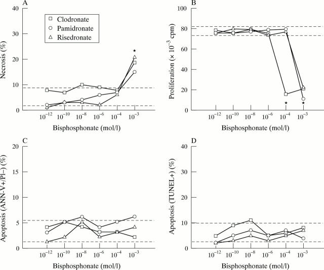
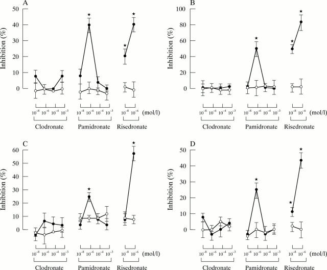
Results: Only high concentrations (>10-6 mol/l) of clodronate, pamidronate, and risedronate induced a decrease in the viability and proliferation of chondrocytes. None of the BP at concentrations ranging from 10-12 to 10-3 mol/l induced apoptosis. Growth retardation and apoptosis induced by dexamethasone (10-7 mol/l) was prevented by addition of pamidronate (10-6 mol/l) or risedronate (10-8 or 10-6 mol/l).
Conclusion: Bisphosphonates in therapeutic concentrations are safe for articular chondrocytes in vitro. Moreover, pamidronate and risedronate prevent dexamethasone-induced growth retardation and apoptosis of chondrocytes. These findings add evidence for a chondroprotective effect of nitrogen-containing BP, especially in patients treated with corticosteroids.
Full Text
The Full Text of this article is available as a PDF (87.4 KB).
Figure 1 .
Influence of clodronate, pamidronate, and risedronate on chondrocyte necrosis (A), proliferation (B), and apoptosis (C/D). (A) Percentage of PI+ necrotic cells (10 000 cells were measured). (B) Proliferation of cells (tests were done in quadruplicate). (C) Percentage of annexin V positive, PI- apoptotic cells (10 000 chondrocytes were measured). (D) Percentage of TUNEL positive apoptotic cells (10 000 cells were measured). Dashed lines represent ranges of control cultures. *p <0.05: chondrocytes incubated with bisphosphonates v control cultures.
Figure 2 .
Influence of clodronate, pamidronate, and risedronate on chondrocytes co-incubated with dexamethasone 10-7 mol/l (closed symbols) or 10-5 mol/l (open symbols). Results expressed as percentage of inhibition of: (A) necrosis; (B) growth retardation; (C) apoptosis measured by annexin V; and (D) apoptosis measured by TUNEL. Bars represent the standard error of the mean of three experiments. *p<0.05: chondrocytes incubated with both dexamethasone and bisphosphonates v chondrocytes incubated with dexamethasone alone.


