Abstract
Objective: To carry out a prospective two year follow up study comparing conventional radiography, three-phase bone scintigraphy, ultrasonography (US), and three dimensional (3D) magnetic resonance imaging (MRI) with precontrast and dynamic postcontrast examination in detecting early arthritis. The aim of the follow up study was to monitor the course of erosions during treatment with disease modifying antirheumatic drugs by different modalities and to determine whether the radiographically occult changes like erosive bone lesions of the finger joints detected by MRI and US in the initial study would show up on conventional radiographs two years later. Additionally, to study the course of soft tissue lesions depicted in the initial study in comparison with the clinical findings.
Methods: The metacarpophalangeal, proximal interphalangeal, and distal interphalangeal joints (14 joints) of the clinically more severely affected hand (soft tissue swelling and joint tenderness) as determined in the initial study of 49 patients with various forms of arthritis were examined twice. The patients had initially been divided into two groups. The follow up group I included 28 subjects (392 joints) without radiographic signs of destructive arthritis (Larsen grades 0–1) of the investigated hand and wrist, and group II (control group) included 21 patients (294 joints) with radiographs showing erosions (Larsen grade 2) of the investigated hand or wrist, or both, at the initial examination.
Results: (1) Radiography at the two year follow up detected only two erosions (two patients) in group I and 10 (nine patients) additional erosions in group II. Initial MRI had already detected both erosions in group I and seven (seven patients) of the 10 erosions in group II. Initial US had depicted one erosion in group I and four of the 10 erosions in group II. (2) In contrast with conventional radiography, 3D MRI and US demonstrated an increase in erosions in comparison with the initial investigation. (3) The abnormal findings detected by scintigraphy were decreased at the two year follow up. (4) Both groups showed a marked clinical improvement of synovitis and tenosynovitis, as also shown by MRI and US. (5) There was a striking discrepancy between the decrease in the soft tissue lesions as demonstrated by clinical findings, MRI, and US, and the significant increase in erosive bone lesions, which were primarily evident at MRI and US.
Conclusions: Despite clinical improvement and a regression of inflammatory soft tissue lesions, erosive bone lesions were increased at the two year follow up, which were more pronounced with 3D MRI and less pronounced with US. The results of our study suggest that owing to the inadequate depiction of erosions and soft tissue lesions, conventional radiography alone has limitations in the intermediate term follow up of treatment. US has a high sensitivity for depicting inflammatory soft tissue lesions, but dynamic 3D MRI is more sensitive in differentiating minute erosions.
Full Text
The Full Text of this article is available as a PDF (367.8 KB).
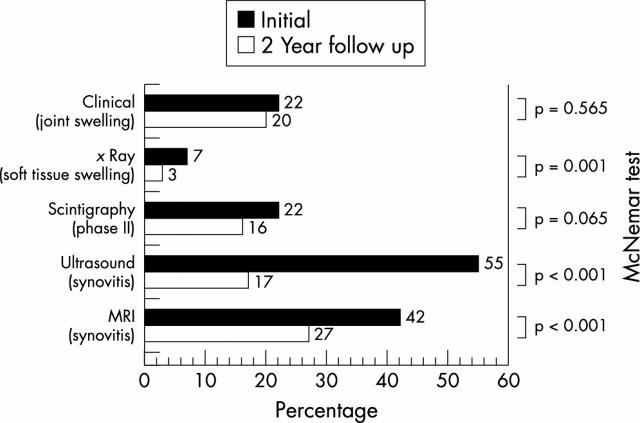
Figure 1 .
Detection of soft tissue lesions (%) by the different modalities in group I (n=392 finger joints = 100%).
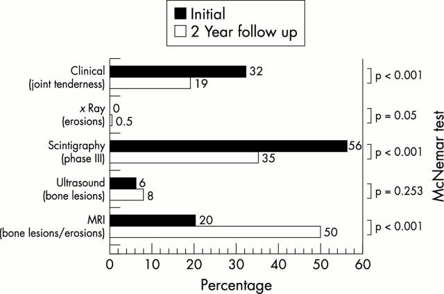
Figure 2 .
Detection of bone lesions (%) by the different modalities in group I (n=392 finger joints = 100%).
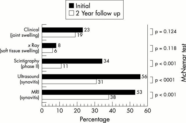
Figure 3 .
Detection of soft tissue lesions (%) by the different modalities in group II (n=294 finger joints = 100%).
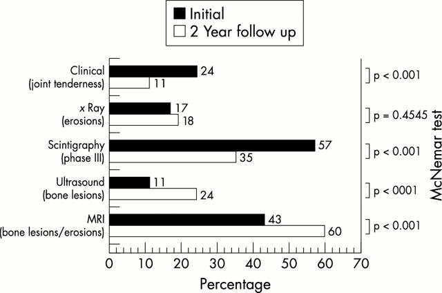
Figure 4 .
Detection of bone lesions (%) by the different modalities in group II (n=294 finger joints = 100%).
Figure 5 .
Images obtained by radiography (A-D), MRI (E-I), scintigraphy (J-M), ultrasound (N-P) of the clinically most severely affected left hand in a woman with RA aged 20 at the time of the initial examination (time 0) and 22 at the time of follow up. (A, B) Survey radiograph of the left hand at time 0 in dorsovolar projection (A) and with the hand in the volardorsal, semisupine (45°) position with abducted fingers, the so-called "zither player position" (B): demonstration of a narrowed joint cleft in PIP joint III and of a small cystoid brightening on the radial side without disruption of the border lamella at the head of the proximal phalanx of digit III (open arrow). There is partial loss of the subchondral border lamella at the ulnar base of the proximal phalanx of digit IV (closed arrow). These changes do not yet represent direct signs of arthritis (Larsen stage 1). (C, D) Follow up radiography of the left hand performed two years after the initial examination in the dorsovolar projection (C) and in the zither player position (D): the joint clefts of all PIP joints now clearly show narrowing. The cystoid brightening at the head of the proximal phalanx of digit III seen at time 0 is now definitely identified as an erosion, but only on the film obtained in the zither player position (thick arrow) and not on the dorsovolar projection. The partial loss of the subchondral border lamella at the ulnar base of the proximal phalanx of digit IV now shows recalcification. Newly developed cystoid lesions not disrupting the border lamellae (thin arrows) are seen on the radial side of the head of the proximal phalanx of digit II, on the ulnar side of the head of the proximal phalanx of digit V, at the metacarpal head of digit I, and in corresponding locations at the scaphotrapezoid joint. The new erosion in PIP joint III is a direct sign of arthritis affecting less than 25% of the joint area, corresponding to Larsen stage 2.
Figure 5 .
(contd) (E, F, G) MRI of the left hand performed at time 0 using an unenhanced T1 weighted 3D gradient echo sequence. The figure only shows one coronal section (E) obtained before contrast administration and a corresponding postcontrast section (F, G). (E) A large erosion on the radial side of the head of the proximal phalanx of digit III (open arrow) can be seen, which is demonstrated by MRI in association with extensive synovitis (asterisk) at PIP III already two years before there is clear cut radiographic evidence of its presence (fig 5D). After contrast medium administration (F), this erosion is partly obscured by the pronounced enhancement of the synovitis (*) at PIP III. Additional, smaller erosions—likewise not demonstrated radiographically—are seen in the metacarpal head of digit IV (arrow with loop) (more clearly seen in other images), on the ulnar side of the head of the proximal phalanx of PIP IV (thin, white arrow), and on the ulnar side of the metacarpal head of digit V (white arrow ) (G). Florid, contrast-enhanced synovitis (*, s) is depicted at PIP joints I–V, and at MCP joints I, II, IV, V. Florid, contrast-enhancing tenosynovitis (t) is seen affecting the flexor tendons of fingers 1–5. The radiographically demonstrated (fig 5A) partial loss of the subchondral border lamella at the ulnar base of the proximal phalanx of digit IV corresponds to contrast-enhancing osteitis affecting the entire base of the phalanx at MRI (black arrow). (H, I) After two years of DMARD treatment the patient underwent follow up MRI of the left hand using the same parameters as at time 0 using a comparable section orientation. The known lesion on the radial side of PIP joint III shows regression but still shows pronounced enhancement reflecting florid activity. The erosion on the ulnar side of PIP joint IV (open arrow (I) shows progression in the presence of more extensive synovitis (*, I). The tenosynovitis on the flexor side of digit III has a higher signal intensity on the postcontrast image than at time 0.
Figure 5 .
(contd) (J, K) Initial scintigraphy, phases II (J) and III (K), shows hot spots in PIP joints II, III, and IV and in the wrist (lunate bone or adjacent parts of ulnar and radial bones). (L, M) At follow up two years later, scintigraphy, phase II (L), shows hot spots in the wrist, MCP joints I and III, IP joint, PIP joints II and III. Scintigraphy, phase III (M), shows hot spots in the wrist, MCP joints I, III, and V, interphalangeal joint, and DIP joints II, III, and IV.
Figure 5 .
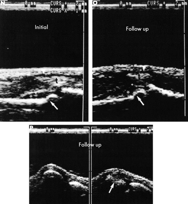
(contd) (N, O, P) (N) The initial ultrasound examination showed synovitis of the proximal interphalangeal (PIP) joints II, III. Ultrasound shows a hypointense line indicating synovitis (*) in PIP joints II (left) and III (right). The flexor tendon sheaths (arrowheads) of the second and third fingers are distended as an indication of tenosynovitis (t, flexor tendon). The step-like interruption of the bone margin (arrow) of the head of the proximal phalanx of digits II and III is indicative of erosion. (O) The figure shows a hypointense line indicating synovitis (*) in PIP joints II (left) and III (right). The flexor tendon sheaths (arrowheads) of the second and third fingers show minimal distention as an indication of tenosynovitis (t, flexor tendon). Irregularities of the joint contour were found at the heads of the proximal phalanx II and III (arrows). Ultrasound shows a large erosion at the head (arrow) of MCP joint V (P, right side) in the dorsal aspect and in the flexed MCP joint, which was not seen at conventional radiography (figs 5A-D) but in both MRI examinations (initially and at the two year follow up, fig 5G). The left side shows a normal joint contour of MCP joint IV.
Selected References
These references are in PubMed. This may not be the complete list of references from this article.
- Alasaarela E., Takalo R., Tervonen O., Hakala M., Suramo I. Sonography and MRI in the evaluation of painful arthritic shoulder. Br J Rheumatol. 1997 Sep;36(9):996–1000. doi: 10.1093/rheumatology/36.9.996. [DOI] [PubMed] [Google Scholar]
- Armour K. J., Armour K. E., van't Hof R. J., Reid D. M., Wei X. Q., Liew F. Y., Ralston S. H. Activation of the inducible nitric oxide synthase pathway contributes to inflammation-induced osteoporosis by suppressing bone formation and causing osteoblast apoptosis. Arthritis Rheum. 2001 Dec;44(12):2790–2796. doi: 10.1002/1529-0131(200112)44:12<2790::aid-art466>3.0.co;2-x. [DOI] [PubMed] [Google Scholar]
- Arnett F. C., Edworthy S. M., Bloch D. A., McShane D. J., Fries J. F., Cooper N. S., Healey L. A., Kaplan S. R., Liang M. H., Luthra H. S. The American Rheumatism Association 1987 revised criteria for the classification of rheumatoid arthritis. Arthritis Rheum. 1988 Mar;31(3):315–324. doi: 10.1002/art.1780310302. [DOI] [PubMed] [Google Scholar]
- Backhaus M., Kamradt T., Sandrock D., Loreck D., Fritz J., Wolf K. J., Raber H., Hamm B., Burmester G. R., Bollow M. Arthritis of the finger joints: a comprehensive approach comparing conventional radiography, scintigraphy, ultrasound, and contrast-enhanced magnetic resonance imaging. Arthritis Rheum. 1999 Jun;42(6):1232–1245. doi: 10.1002/1529-0131(199906)42:6<1232::AID-ANR21>3.0.CO;2-3. [DOI] [PubMed] [Google Scholar]
- Bollow M., Biedermann T., Kannenberg J., Paris S., Schauer-Petrowski C., Minden K., Schöntube M., Hamm B., Sieper J., Braun J. Use of dynamic magnetic resonance imaging to detect sacroiliitis in HLA-B27 positive and negative children with juvenile arthritides. J Rheumatol. 1998 Mar;25(3):556–564. [PubMed] [Google Scholar]
- Bollow M., Braun J., Hamm B., Eggens U., Schilling A., König H., Wolf K. J. Early sacroiliitis in patients with spondyloarthropathy: evaluation with dynamic gadolinium-enhanced MR imaging. Radiology. 1995 Feb;194(2):529–536. doi: 10.1148/radiology.194.2.7824736. [DOI] [PubMed] [Google Scholar]
- Braun J., Bollow M., Eggens U., König H., Distler A., Sieper J. Use of dynamic magnetic resonance imaging with fast imaging in the detection of early and advanced sacroiliitis in spondylarthropathy patients. Arthritis Rheum. 1994 Jul;37(7):1039–1045. doi: 10.1002/art.1780370709. [DOI] [PubMed] [Google Scholar]
- Braun J., Bollow M., Seyrekbasan F., Häberle H. J., Eggens U., Mertz A., Distler A., Sieper J. Computed tomography guided corticosteroid injection of the sacroiliac joint in patients with spondyloarthropathy with sacroiliitis: clinical outcome and followup by dynamic magnetic resonance imaging. J Rheumatol. 1996 Apr;23(4):659–664. [PubMed] [Google Scholar]
- Brower A. C. Use of the radiograph to measure the course of rheumatoid arthritis. The gold standard versus fool's gold. Arthritis Rheum. 1990 Mar;33(3):316–324. doi: 10.1002/art.1780330303. [DOI] [PubMed] [Google Scholar]
- Conaghan P. G., Brooks P. Disease-modifying antirheumatic drugs, including methotrexate, gold, antimalarials, and D-penicillamine. Curr Opin Rheumatol. 1995 May;7(3):167–173. doi: 10.1097/00002281-199505000-00003. [DOI] [PubMed] [Google Scholar]
- Dawes P. T., Fowler P. D. Treatment of early rheumatoid arthritis: a review of current and future concepts and therapy. Clin Exp Rheumatol. 1995 May-Jun;13(3):381–394. [PubMed] [Google Scholar]
- Devlin J., Lilley J., Gough A., Huissoon A., Holder R., Reece R., Perkins P., Emery P. Clinical associations of dual-energy X-ray absorptiometry measurement of hand bone mass in rheumatoid arthritis. Br J Rheumatol. 1996 Dec;35(12):1256–1262. doi: 10.1093/rheumatology/35.12.1256. [DOI] [PubMed] [Google Scholar]
- Dougados M., van der Linden S., Juhlin R., Huitfeldt B., Amor B., Calin A., Cats A., Dijkmans B., Olivieri I., Pasero G. The European Spondylarthropathy Study Group preliminary criteria for the classification of spondylarthropathy. Arthritis Rheum. 1991 Oct;34(10):1218–1227. doi: 10.1002/art.1780341003. [DOI] [PubMed] [Google Scholar]
- Emery P. Early rheumatoid arthritis: therapeutic strategies. Scand J Rheumatol Suppl. 1994;100:3–7. doi: 10.3109/03009749409095196. [DOI] [PubMed] [Google Scholar]
- Foley-Nolan D., Stack J. P., Ryan M., Redmond U., Barry C., Ennis J., Coughlan R. J. Magnetic resonance imaging in the assessment of rheumatoid arthritis--a comparison with plain film radiographs. Br J Rheumatol. 1991 Apr;30(2):101–106. doi: 10.1093/rheumatology/30.2.101. [DOI] [PubMed] [Google Scholar]
- Forslind K., Larsson E. M., Johansson A., Svensson B. Detection of joint pathology by magnetic resonance imaging in patients with early rheumatoid arthritis. Br J Rheumatol. 1997 Jun;36(6):683–688. doi: 10.1093/rheumatology/36.6.683. [DOI] [PubMed] [Google Scholar]
- Gaffney K., Cookson J., Blake D., Coumbe A., Blades S. Quantification of rheumatoid synovitis by magnetic resonance imaging. Arthritis Rheum. 1995 Nov;38(11):1610–1617. doi: 10.1002/art.1780381113. [DOI] [PubMed] [Google Scholar]
- Gilkeson G., Polisson R., Sinclair H., Vogler J., Rice J., Caldwell D., Spritzer C., Martinez S. Early detection of carpal erosions in patients with rheumatoid arthritis: a pilot study of magnetic resonance imaging. J Rheumatol. 1988 Sep;15(9):1361–1366. [PubMed] [Google Scholar]
- Gough A. K., Lilley J., Eyre S., Holder R. L., Emery P. Generalised bone loss in patients with early rheumatoid arthritis. Lancet. 1994 Jul 2;344(8914):23–27. doi: 10.1016/s0140-6736(94)91049-9. [DOI] [PubMed] [Google Scholar]
- Grassi W., Filippucci E., Farina A., Salaffi F., Cervini C. Ultrasonography in the evaluation of bone erosions. Ann Rheum Dis. 2001 Feb;60(2):98–103. doi: 10.1136/ard.60.2.98. [DOI] [PMC free article] [PubMed] [Google Scholar]
- Hartley R. M., Liang M. H., Weissman B. N., Sosman J. L., Katz R., Charlton J. R. The value of conventional views and radiographic magnification in evaluating early rheumatoid arthritis. Arthritis Rheum. 1984 Jul;27(7):744–751. doi: 10.1002/art.1780270704. [DOI] [PubMed] [Google Scholar]
- Imhoff A. B., Hodler J. Correlation of MR imaging, CT arthrography, and arthroscopy of the shoulder. Bull Hosp Jt Dis. 1996;54(3):146–152. [PubMed] [Google Scholar]
- Jorgensen C., Cyteval C., Anaya J. M., Baron M. P., Lamarque J. L., Sany J. Sensitivity of magnetic resonance imaging of the wrist in very early rheumatoid arthritis. Clin Exp Rheumatol. 1993 Mar-Apr;11(2):163–168. [PubMed] [Google Scholar]
- Kieft G. J., Dijkmans B. A., Bloem J. L., Kroon H. M. Magnetic resonance imaging of the shoulder in patients with rheumatoid arthritis. Ann Rheum Dis. 1990 Jan;49(1):7–11. doi: 10.1136/ard.49.1.7. [DOI] [PMC free article] [PubMed] [Google Scholar]
- Koenig H., Lucas D., Meissner R. The wrist: a preliminary report on high-resolution MR imaging. Radiology. 1986 Aug;160(2):463–467. doi: 10.1148/radiology.160.2.3726128. [DOI] [PubMed] [Google Scholar]
- Larsen A., Dale K., Eek M. Radiographic evaluation of rheumatoid arthritis and related conditions by standard reference films. Acta Radiol Diagn (Stockh) 1977 Jul;18(4):481–491. doi: 10.1177/028418517701800415. [DOI] [PubMed] [Google Scholar]
- Maini R. N., Breedveld F. C., Kalden J. R., Smolen J. S., Davis D., Macfarlane J. D., Antoni C., Leeb B., Elliott M. J., Woody J. N. Therapeutic efficacy of multiple intravenous infusions of anti-tumor necrosis factor alpha monoclonal antibody combined with low-dose weekly methotrexate in rheumatoid arthritis. Arthritis Rheum. 1998 Sep;41(9):1552–1563. doi: 10.1002/1529-0131(199809)41:9<1552::AID-ART5>3.0.CO;2-W. [DOI] [PubMed] [Google Scholar]
- Manger B., Kalden J. R. Joint and connective tissue ultrasonography--a rheumatologic bedside procedure? A German experience. Arthritis Rheum. 1995 Jun;38(6):736–742. doi: 10.1002/art.1780380603. [DOI] [PubMed] [Google Scholar]
- McGonagle D., Conaghan P. G., O'Connor P., Gibbon W., Green M., Wakefield R., Ridgway J., Emery P. The relationship between synovitis and bone changes in early untreated rheumatoid arthritis: a controlled magnetic resonance imaging study. Arthritis Rheum. 1999 Aug;42(8):1706–1711. doi: 10.1002/1529-0131(199908)42:8<1706::AID-ANR20>3.0.CO;2-Z. [DOI] [PubMed] [Google Scholar]
- McQueen F. M., Stewart N., Crabbe J., Robinson E., Yeoman S., Tan P. L., McLean L. Magnetic resonance imaging of the wrist in early rheumatoid arthritis reveals a high prevalence of erosions at four months after symptom onset. Ann Rheum Dis. 1998 Jun;57(6):350–356. doi: 10.1136/ard.57.6.350. [DOI] [PMC free article] [PubMed] [Google Scholar]
- McQueen F. M., Stewart N., Crabbe J., Robinson E., Yeoman S., Tan P. L., McLean L. Magnetic resonance imaging of the wrist in early rheumatoid arthritis reveals progression of erosions despite clinical improvement. Ann Rheum Dis. 1999 Mar;58(3):156–163. doi: 10.1136/ard.58.3.156. [DOI] [PMC free article] [PubMed] [Google Scholar]
- Moreland L. W., Baumgartner S. W., Schiff M. H., Tindall E. A., Fleischmann R. M., Weaver A. L., Ettlinger R. E., Cohen S., Koopman W. J., Mohler K. Treatment of rheumatoid arthritis with a recombinant human tumor necrosis factor receptor (p75)-Fc fusion protein. N Engl J Med. 1997 Jul 17;337(3):141–147. doi: 10.1056/NEJM199707173370301. [DOI] [PubMed] [Google Scholar]
- Moreland L. W., Daniel W. W., Alarcón G. S. The value of the Nørgaard view in the evaluation of erosive arthritis. J Rheumatol. 1990 May;17(5):614–617. [PubMed] [Google Scholar]
- Munk P. L., Vellet A. D., Levin M. F., Bell D. A., Harth M. M., McCain G. A. Intravenous administration of gadolinium in the evaluation of rheumatoid arthritis of the shoulder. Can Assoc Radiol J. 1993 Apr;44(2):99–106. [PubMed] [Google Scholar]
- NORGAARD F. EARLIEST ROENTGENOLOGICAL CHANGES IN POLYARTHRITIS OF THE RHEUMATOID TYPE: RHEUMATOID ARTHRITIS. Radiology. 1965 Aug;85:325–329. doi: 10.1148/85.2.325. [DOI] [PubMed] [Google Scholar]
- Ostergaard M., Hansen M., Stoltenberg M., Gideon P., Klarlund M., Jensen K. E., Lorenzen I. Magnetic resonance imaging-determined synovial membrane volume as a marker of disease activity and a predictor of progressive joint destruction in the wrists of patients with rheumatoid arthritis. Arthritis Rheum. 1999 May;42(5):918–929. doi: 10.1002/1529-0131(199905)42:5<918::AID-ANR10>3.0.CO;2-2. [DOI] [PubMed] [Google Scholar]
- Ostergaard M. MRI bone erosions and MRI bone lesions in early rheumatoid arthritis: comment on the article by McGonagle et al. Arthritis Rheum. 2000 Apr;43(4):949–950. doi: 10.1002/1529-0131(200004)43:4<949::AID-ANR34>3.0.CO;2-6. [DOI] [PubMed] [Google Scholar]
- Ostergaard M., Stoltenberg M., Gideon P., Sørensen K., Henriksen O., Lorenzen I. Changes in synovial membrane and joint effusion volumes after intraarticular methylprednisolone. Quantitative assessment of inflammatory and destructive changes in arthritis by MRI. J Rheumatol. 1996 Jul;23(7):1151–1161. [PubMed] [Google Scholar]
- Pierre-Jerome C., Bekkelund S. I., Husby G., Mellgren S. I., Torbergsen T. Bilateral fast MR imaging of the rheumatoid wrist. Clin Rheumatol. 1996 Jan;15(1):42–46. doi: 10.1007/BF02231683. [DOI] [PubMed] [Google Scholar]
- Pierre-Jerome C., Bekkelund S. I., Mellgren S. I., Torbergsen T., Husby G., Nordstrøm R. The rheumatoid wrist: bilateral MR analysis of the distribution of rheumatoid lesions in axial plan in a female population. Clin Rheumatol. 1997 Jan;16(1):80–86. doi: 10.1007/BF02238768. [DOI] [PubMed] [Google Scholar]
- Poleksic L., Zdravkovic D., Jablanovic D., Watt I., Bacic G. Magnetic resonance imaging of bone destruction in rheumatoid arthritis: comparison with radiography. Skeletal Radiol. 1993 Nov;22(8):577–580. doi: 10.1007/BF00197138. [DOI] [PubMed] [Google Scholar]
- Rau R., Herborn G. A modified version of Larsen's scoring method to assess radiologic changes in rheumatoid arthritis. J Rheumatol. 1995 Oct;22(10):1976–1982. [PubMed] [Google Scholar]
- Tan E. M., Cohen A. S., Fries J. F., Masi A. T., McShane D. J., Rothfield N. F., Schaller J. G., Talal N., Winchester R. J. The 1982 revised criteria for the classification of systemic lupus erythematosus. Arthritis Rheum. 1982 Nov;25(11):1271–1277. doi: 10.1002/art.1780251101. [DOI] [PubMed] [Google Scholar]
- Tonolli-Serabian I., Poet J. L., Dufour M., Carasset S., Mattei J. P., Roux H. Magnetic resonance imaging of the wrist in rheumatoid arthritis: comparison with other inflammatory joint diseases and control subjects. Clin Rheumatol. 1996 Mar;15(2):137–142. doi: 10.1007/BF02230330. [DOI] [PubMed] [Google Scholar]
- Vestring T., Bongartz G., Konermann W., Erlemann R., Reuther G., Krings W., Saathoff J., Drescher H., Peters P. E. Stellenwert der Magnetresonanztomographie in der Diagnostik von Schultergelenkerkrankungen. Rofo. 1991 Feb;154(2):143–149. doi: 10.1055/s-2008-1033102. [DOI] [PubMed] [Google Scholar]
- Wakefield R. J., Gibbon W. W., Conaghan P. G., O'Connor P., McGonagle D., Pease C., Green M. J., Veale D. J., Isaacs J. D., Emery P. The value of sonography in the detection of bone erosions in patients with rheumatoid arthritis: a comparison with conventional radiography. Arthritis Rheum. 2000 Dec;43(12):2762–2770. doi: 10.1002/1529-0131(200012)43:12<2762::AID-ANR16>3.0.CO;2-#. [DOI] [PubMed] [Google Scholar]
- Weiss K. L., Beltran J., Lubbers L. M. High-field MR surface-coil imaging of the hand and wrist. Part II. Pathologic correlations and clinical relevance. Radiology. 1986 Jul;160(1):147–152. doi: 10.1148/radiology.160.1.3715026. [DOI] [PubMed] [Google Scholar]
- Wilke W. S., Sweeney T. J., Calabrese L. H. Early, aggressive therapy for rheumatoid arthritis: concerns, descriptions, and estimate of outcome. Semin Arthritis Rheum. 1993 Oct;23(2 Suppl 1):26–41. doi: 10.1016/s0049-0172(10)80005-8. [DOI] [PubMed] [Google Scholar]
Associated Data
This section collects any data citations, data availability statements, or supplementary materials included in this article.


