

Full Text
The Full Text of this article is available as a PDF (122.0 KB).
Figure 1 .
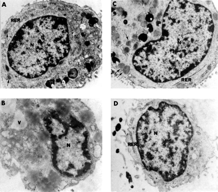
Transmission electron micrographs of chondrocytes cultured in vitro for 72 hours. (A) Basal conditions: the nucleus (N) appears euchromatic, the cytoplasm contains abundant rough endoplasmic reticulum (RER) and lipid droplets (arrows). x9000. (B) IL1ß present: the cell shows a vacuolate cytoplasm devoid of the typical structure. N, nucleus; V, vacuole. x9000. (C) Basal conditions and given PST stimulation: the cell shows an euchromatic nucleus (N) and a cytoplasm rich in rough endoplasmic reticulum (RER) and lipid droplets (arrows). x10 000. (D) IL1ß present and given PST stimulation: the cell clearly recovers its good state of health. Rough endoplasmic reticulum (RER) is present and vacuoles are almost absent. N, nucleus. x9000.