Abstract
Objective: To investigate the relationship between these conditions and bone mineral density (BMD) in Indo-Asian rheumatology patients.
Methods: 110 Indo-Asian patients presenting to a general rheumatology clinic were selected for the study, of whom 77 were vitamin D deficient (HD), and 33 also had associated secondary hyperparathyroidism (HD-SHPT). BMD measurements at the femoral neck (FN), lumbar spine (LS), distal radius (DR), and radius 33% were obtained. T and Z scores were used in the evaluation of the dual energy x ray absorptiometry findings. The Mann-Whitney U test and χ2 tests were used in the statistical analysis.
Results: The patients with HD-SHPT had significantly lower serum 25-OH-vitamin D, higher serum alkaline phosphatase, and lower calcium levels than the HD group. Both T and Z scores failed to show any significant difference between the groups in the LS, but patients with HD-SHPT compared with those with HD had significantly lower FN T scores (-1.05 (1.47) v 0.28 (1.22), p=0.003), FN Z scores (-0.45 (1.27) v 0.26 (0.96), p=0.001), DR T scores (-1.83 (1.82) v -0.44 (1.77), p<0.001), DR Z scores (-1.25 (1.44) v –0.08 (1.6), p=0.001), radius 33% T scores (-1.65 (1.66) v –0.79 (1.21), p=0.008), and radius 33% Z scores (-1.07 (1.28) v –0.44 (1.1), p=0.017).
Conclusion: Hypovitaminosis D complicated by secondary hyperparathyroidism is associated with significantly decreased bone mineral density.
Full Text
The Full Text of this article is available as a PDF (94.4 KB).
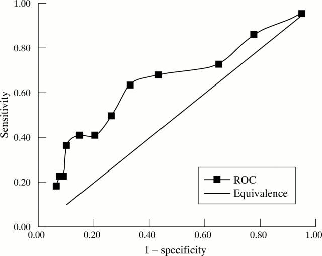
Figure 1 .
Receiver operator curve in which osteoporosis is defined as a distal radius T score of <-2.5, for different PTH cut off points.
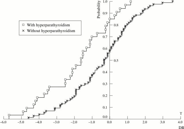
Figure 2 .
Cumulative probability plots for distal radius bone mineral density T score distribution among Indo-Asian rheumatology patients with hypovitaminosis D associated with hyperparathyroidism (n=33), compared with those without hyperparathyroidism (n=77).


