

Full Text
The Full Text of this article is available as a PDF (180.0 KB).
Figure 1 .
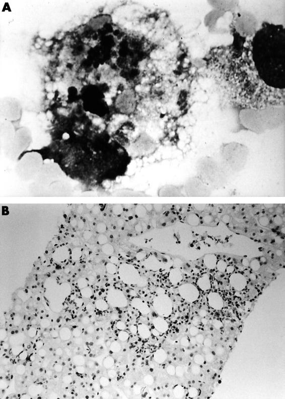
Stomach of a patient with lSSc—muscle coat. (A) Circular and longitudinal muscle layers. Note the marked fibrosis among the smooth muscle cells (smcs) (arrows). Sometimes smcs show a severe cytoplasmic vacuolisation (arrowheads); (B) a group of smcs. Myofilaments and dense bodies show a severe disarray (arrows). The dense bodies are thickened (arrowheads). F, collagen fibres; (C) nerve bundle containing many axons in myenteric plexus area. The axoplasm appears oedematous and poor in neurotubules and neurofilaments (arrowheads). L, lipofuscinic bodies; (D) nerve ending (NE), embedded in abundant collagen fibres (F) shows just one close contact area with an smc (arrows); (E) blood vessel, with lumen completely occluded by erythrocytes (E) and neutrophils (N) is close to a nerve bundle (Nb) containing many altered axons; (F) mast cell rich in granules is evident between a blood vessel (BV) and the smcs. Original magnification: (A) x5000; (B, E, F) x3000; (C) x8000; (D) x6000.