

Abstract
Methods: HAC in vitro were incubated with and without histamine in 96 well culture plates and the extent of cell proliferation was determined using the naphthol blue-black method. Histamine effects were analysed with the histamine H1 and H2 receptor antagonists, mepyramine and ranitidine, respectively. Rabbit polyclonal antibodies and alkaline phosphatase conjugated secondary antibodies were used, and histamine and HDC were demonstrated by immunohistochemistry in OA cartilage tissues.
Results: Histamine stimulated the proliferation of HAC in culture. This stimulation was blocked by the addition of mepyramine, but not ranitidine, suggesting that the effect is mediated through H1 histamine receptors. The addition of α-fluoromethylhistidine, a specific inhibitor of histidine decarboxylase (the enzyme responsible for histamine production), reduced the rate of proliferation of HAC. Both histamine and histidine decarboxylase were demonstrated in chondrocytes of OA cartilage by immunohistochemistry.
Conclusion: Changes induced by histamine in the proliferative rate of HAC may contribute to the formation of chondrocyte clusters associated with OA cartilage; an observation supported by the demonstration of histamine and HDC expression by chondrocytes of OA cartilage in situ.
Full Text
The Full Text of this article is available as a PDF (139.2 KB).
Figure 1.
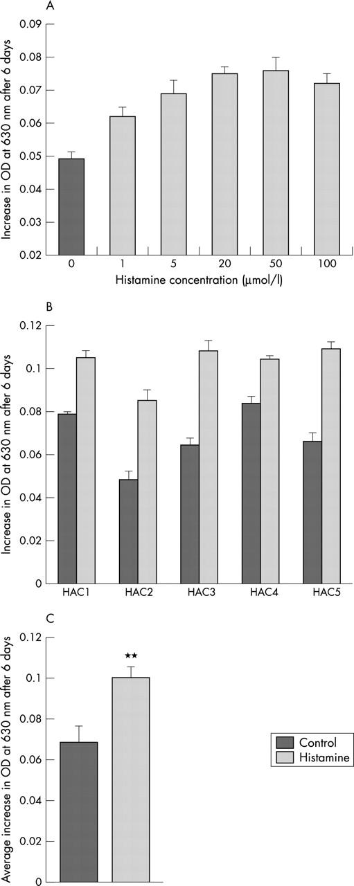
Effect of histamine on the proliferative rate of HAC in vitro. (A) Effect of different histamine concentrations (1–100 µmol/l) on cell proliferation after six days. Preparation HAC2, passage 2. Data represent intra-assay values, showing mean (SEM) of eight wells for each treatment. (B) Effect of histamine (20 µmol/l) on cell proliferation of five different HAC cultures after six days. Data represent intra-assay values, showing mean (SEM) of eight wells for each treatment. (C) Averaged data from five HAC cultures with or without histamine (20 µmol/l). Data represent the mean (SEM) of the five HAC cultures presented in (B). **p=0.00012, Student's t test.
Figure 2.
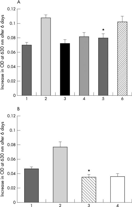
(A) Effect of specific H1 and H2 histamine receptor antagonists on the histamine stimulated proliferation of HAC. 1, Control culture; 2, + histamine (20 µmol/l); 3, + mepyramine (20 µmol/l); 4, + ranitidine (20 µmol/l); 5, + histamine (20 µmol/l) and mepyramine (20 µmol/l); 6, + histamine (20 µmol/l) and ranitidine (20 µmol/l). Note the significant inhibition of the histamine effect by mepyramine (*p=0.003, Student's t test). Representative experiment (n=3) using preparation HAC3, passage 2, this being qualitatively similar for HAC2 and 5. (B) Effect of α-FMH on the proliferation of HAC. 1, Control culture; 2, + histamine (20 µmol/l); 3, + α-FMH (100 µmol/l); 4, + α-FMH (100 µmol/l) and mepyramine (20 µmol/l). Representative experiment showing qualitatively similar data from two HAC cultures. Note modest reduction in proliferative rate of HAC by α-FMH (*p=0.05, Student's t test).
Figure 3.
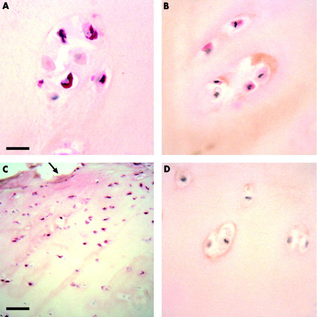
Immunolocalisation of histamine and histidine decarboxylase in chondrocytes of OA cartilage. (A) Tissue section of OA cartilage showing a proportion of the cells in a chondrocyte cluster positive for histamine (red). (B) Tissue section of OA cartilage with a proportion of cells of a chondrocyte cluster immunostained for HDC (red). (C) Low power micrograph of tissue section of OA cartilage showing histamine expression by HAC predominantly in the superficial articular surface (arrowed), and not in the deeper regions of the articular cartilage. (D) Control tissue section with normal rabbit IgG as substitute for primary antibodies showing no immunostaining. Bar = 25 µm for micrographs A, B, and D; bar = 140 µm for micrograph C. All sections were lightly counterstained with Harris's haematoxylin.