

Abstract
Objective: To analyse the early changes leading to OA by examining the articular cytokine expression and degenerative changes in STR/ort mice.
Methods: 122 STR/ort mice of both sexes aged between 2 and 15.5 months were included. Thin sections of the knees were analysed for osteoarthritic changes by haematoxylin/eosin staining. The articular cytokine expression was investigated by immunohistochemical staining using monoclonal antibodies specific for interleukin (IL)6, tumour necrosis factor α, transforming growth factor ß1 (TGFß1), IL1ß, IL4, and IL10, respectively.
Results: Both cartilage degeneration and articular cytokine expression differ between the sexes. The protection from cartilage degeneration in the female mice correlates with an increased expression of TGFß1 and IL4 at 2 months of age.
Conclusion: The increased expression of TGFß1 and IL4 in young STR/ort female mice suggests that the sexual dimorphism is mediated through the articular expression of cytokines involved in cartilage metabolism.
Full Text
The Full Text of this article is available as a PDF (238.4 KB).
Figure 1 .

Male and female STR/ort mice differ in the severity of OA and in the articular cytokine expression. Cartilage degradation and cytokine expression pattern from male (left panels) and female mice (right panels) are shown. Data from STR/ort mice are shown as filled symbols, data from BALB/c mice are shown as open symbols to the left and to the right of the STR/ort data, respectively. (A) Degradation of the articular cartilage. Squares (males) and circles (females) resemble the course of degenerative changes and show the mean grades of cartilage degradation. (B) Cytokine expression patterns of the articular chondrocytes. The medians of the percentages of chondrocytes stained positive for the expression of IL1ß, IL4, and TGFß1 are shown.
Figure 2 .
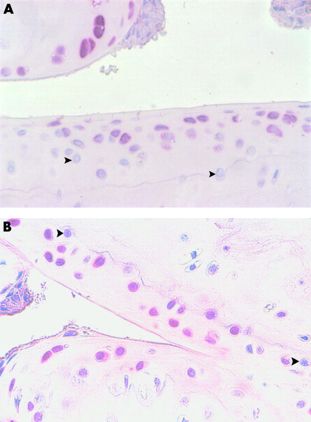
Cytokine expression in the articular cartilage of STR/ort mice. Thin sections of the knees of STR/ort mice were stained immunohistochemically for the presence of the different cytokines. Six hundredfold magnifications of thin sections stained for IL1ß (A) and TGFß1 (B) are shown. Examples for cytokine negative chondrocytes are indicated by black arrowheads.