

Abstract
Objectives: To develop an objective method of nailfold capillaroscopy (NFC), applicable to a wide age range of paediatric patients. To compare the morphological characteristics of the nailfold capillaries in different rheumatology patient groups and controls.
Methods: A colour digital video camera attached to a stereomicroscope was used to capture nailfold capillary images. Computerised image processing was used to analyse and store data. Subsequent quantitative and qualitative morphological analysis was performed in the following paediatric patient and control groups: 18 children with connective tissue diseases (CTD: juvenile dermatomyositis, systemic sclerosis, and undifferentiated connective tissue disease), eight with systemic lupus erythematosus, nine with primary Raynaud's disease, three with primary vasculitis, 15 with juvenile idiopathic arthritis, 17 healthy children and 20 healthy adults. Images were analysed by a single assessor who was unaware of the patient details.
Results: The NFC technique was simple to perform and gave reproducible results, although some intra- and intersubject variation was noted. Capillary density and width was age related, with younger children having fewer and wider capillaries than older children and adults. Linear capillary density was significantly higher in healthy adults (mean (SD) 8.6 (1.6) capillaries/mm) compared with healthy children (HC 6.9 (0.9) capillaries/mm). The group with CTD had the most abnormal findings, with lower linear density (4.9 (1.7) capillaries/mm) and increased capillary loop width (10.7 (7.3) mm) compared with HC (3.5 (1.7) mm). In addition, 11/18 (61%) patients in the CTD group had more than two definitely abnormal capillaries in at least two nailfolds, an abnormality not seen in other subjects. Two qualitative measures, the degree of avascularity and general disarrangement of capillary pattern, were more commonly observed in the CTD group than in HC. The proportion of tortuous capillaries did not differ significantly between study groups.
Conclusions: This study is unique in measuring objective quantitative and qualitative parameters of the nailfold vasculature across a wide spectrum of age and disease. Differences in capillary morphology and frequency in children with CTD compared with other paediatric diseases and healthy controls were demonstrated. In the clinical situation, an assessment of the general degree of disarrangement may offer a fast tool for assessment of the nailfold vasculature which correlates well with NFC data.
Full Text
The Full Text of this article is available as a PDF (173.3 KB).
Figure 1.
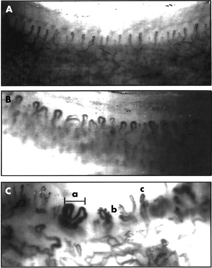
Nailfold capillary morphology: (A) normal capillaries, healthy adult; (B) normal capillaries, healthy 2 year old child; (C) abnormal capillaries, 15 year old boy with JDMS. a, Capillary width in a giant capillary; b, tortuous loop, c, avascular area.
Figure 2.

Linear capillary density. Capillaries/mm—number of capillaries per 1 mm of end capillary row. Healthy adults had significantly increased capillary density compared with healthy children (p<0.01). Patients with CTD also had significantly reduced capillary density compared with healthy children (p<0.001).
Figure 3.

Variation of linear capillary density with age. In a normal population, linear capillary density varies directly with age.
Figure 4.

Capillary loop width in health and disease. mm screen—mean width of the three widest capillaries in millimetres of the screen. 1 mm screen = 15.15 actual µm. Children with CTD had wider capillaries than normal control children (p<0.05).
Selected References
These references are in PubMed. This may not be the complete list of references from this article.
- Andrade L. E., Gabriel Júnior A., Assad R. L., Ferrari A. J., Atra E. Panoramic nailfold capillaroscopy: a new reading method and normal range. Semin Arthritis Rheum. 1990 Aug;20(1):21–31. doi: 10.1016/0049-0172(90)90091-s. [DOI] [PubMed] [Google Scholar]
- Bongard O., Bounameaux H. Clinical investigation of skin microcirculation. Dermatology. 1993;186(1):6–11. doi: 10.1159/000247295. [DOI] [PubMed] [Google Scholar]
- Cony M., Klene-Boudard C., Fontan I., Sanciaume C., Sarrat P., Taieb A., Maleville J. Etude des aspects capillaroscopiques péri-unguéaux chez l'enfant normal. Arch Fr Pediatr. 1992 Mar;49(3):171–174. [PubMed] [Google Scholar]
- Cutolo M., Sulli A., Pizzorni C., Accardo S. Nailfold videocapillaroscopy assessment of microvascular damage in systemic sclerosis. J Rheumatol. 2000 Jan;27(1):155–160. [PubMed] [Google Scholar]
- Duffy C. M., Laxer R. M., Lee P., Ramsay C., Fritzler M., Silverman E. D. Raynaud syndrome in childhood. J Pediatr. 1989 Jan;114(1):73–78. doi: 10.1016/s0022-3476(89)80604-3. [DOI] [PubMed] [Google Scholar]
- Harper F. E., Maricq H. R., Turner R. E., Lidman R. W., Leroy E. C. A prospective study of Raynaud phenomenon and early connective tissue disease. A five-year report. Am J Med. 1982 Jun;72(6):883–888. doi: 10.1016/0002-9343(82)90846-4. [DOI] [PubMed] [Google Scholar]
- Herrick A. L., Moore T., Hollis S., Jayson M. I. The influence of age on nailfold capillary dimensions in childhood. J Rheumatol. 2000 Mar;27(3):797–800. [PubMed] [Google Scholar]
- Houtman P. M., Kallenberg C. G., Fidler V., Wouda A. A. Diagnostic significance of nailfold capillary patterns in patients with Raynaud's phenomenon. An analysis of patterns discriminating patients with and without connective tissue disease. J Rheumatol. 1986 Jun;13(3):556–563. [PubMed] [Google Scholar]
- Houtman P. M., Kallenberg C. G., Wouda A. A., The T. H. Decreased nailfold capillary density in Raynaud's phenomenon: a reflection of immunologically mediated local and systemic vascular disease? Ann Rheum Dis. 1985 Sep;44(9):603–609. doi: 10.1136/ard.44.9.603. [DOI] [PMC free article] [PubMed] [Google Scholar]
- Jayanetti S., Smith C. P., Moore T., Jayson M. I., Herrick A. L. Thermography and nailfold capillaroscopy as noninvasive measures of circulation in children with Raynaud's phenomenon. J Rheumatol. 1998 May;25(5):997–999. [PubMed] [Google Scholar]
- Joyal F., Choquette D., Roussin A., Levington C., Senécal J. L. Evaluation of the severity of systemic sclerosis by nailfold capillary microscopy in 112 patients. Angiology. 1992 Mar;43(3 Pt 1):203–210. doi: 10.1177/000331979204300305. [DOI] [PubMed] [Google Scholar]
- Kabasakal Y., Elvins D. M., Ring E. F., McHugh N. J. Quantitative nailfold capillaroscopy findings in a population with connective tissue disease and in normal healthy controls. Ann Rheum Dis. 1996 Aug;55(8):507–512. doi: 10.1136/ard.55.8.507. [DOI] [PMC free article] [PubMed] [Google Scholar]
- Lee P., Leung F. Y., Alderdice C., Armstrong S. K. Nailfold capillary microscopy in the connective tissue diseases: a semiquantitative assessment. J Rheumatol. 1983 Dec;10(6):930–938. [PubMed] [Google Scholar]
- Lovy M., MacCarter D., Steigerwald J. C. Relationship between nailfold capillary abnormalities and organ involvement in systemic sclerosis. Arthritis Rheum. 1985 May;28(5):496–501. doi: 10.1002/art.1780280505. [DOI] [PubMed] [Google Scholar]
- Maricq H. R. Comparison of quantitative and semiquantitative estimates of nailfold capillary abnormalities in scleroderma spectrum disorders. Microvasc Res. 1986 Sep;32(2):271–276. doi: 10.1016/0026-2862(86)90062-2. [DOI] [PubMed] [Google Scholar]
- Maricq H. R., LeRoy E. C., D'Angelo W. A., Medsger T. A., Jr, Rodnan G. P., Sharp G. C., Wolfe J. F. Diagnostic potential of in vivo capillary microscopy in scleroderma and related disorders. Arthritis Rheum. 1980 Feb;23(2):183–189. doi: 10.1002/art.1780230208. [DOI] [PubMed] [Google Scholar]
- Maricq H. R., Spencer-Green G., LeRoy E. C. Skin capillary abnormalities as indicators of organ involvement in scleroderma (systemic sclerosis), Raynaud's syndrome and dermatomyositis. Am J Med. 1976 Dec;61(6):862–870. doi: 10.1016/0002-9343(76)90410-1. [DOI] [PubMed] [Google Scholar]
- Maricq H. R., Weinberger A. B., LeRoy E. C. Early detection of scleroderma-spectrum disorders by in vivo capillary microscopy: a prospective study of patients with Raynaud's phenomenon. J Rheumatol. 1982 Mar-Apr;9(2):289–291. [PubMed] [Google Scholar]
- Maricq H. R. Wide-field capillary microscopy. Arthritis Rheum. 1981 Sep;24(9):1159–1165. doi: 10.1002/art.1780240907. [DOI] [PubMed] [Google Scholar]
- Martino F., Agolini D., Aprigliano D., Guido F., Placanica G., Giardini O. La capillaroscopia periungueale in soggetti normali di età compresa tra 0 e 16 anni. Minerva Pediatr. 1997 May;49(5):197–201. [PubMed] [Google Scholar]
- Navon P., Yarom A., Davis E. Raynaud's features in childhood. Clinical, immunological and capillaroscopic study. J Mal Vasc. 1992;17(4):273–276. [PubMed] [Google Scholar]
- Nussbaum A. I., Silver R. M., Maricq H. R. Serial changes in nailfold capillary morphology in childhood dermatomyositis. Arthritis Rheum. 1983 Sep;26(9):1169–1172. doi: 10.1002/art.1780260919. [DOI] [PubMed] [Google Scholar]
- Redisch W., Messina E. J., Hughes G., McEwen C. Capillaroscopic observations in rheumatic diseases. Ann Rheum Dis. 1970 May;29(3):244–253. doi: 10.1136/ard.29.3.244. [DOI] [PMC free article] [PubMed] [Google Scholar]
- Rouen L. R., Terry E. N., Doft B. H., Clauss R. H., Redisch W. Classification and measurement of surface microvessels in man. Microvasc Res. 1972 Jul;4(3):285–292. doi: 10.1016/0026-2862(72)90040-4. [DOI] [PubMed] [Google Scholar]
- Scheja A., Akesson A., Niewierowicz I., Wallin L., Wildt M., Wollheim F. A. Computer based quantitative analysis of capillary abnormalities in systemic sclerosis and its relation to plasma concentration of von Willebrand factor. Ann Rheum Dis. 1996 Jan;55(1):52–56. doi: 10.1136/ard.55.1.52. [DOI] [PMC free article] [PubMed] [Google Scholar]
- Scheja A., Elborgh R., Wildt M. Decreased capillary density in juvenile dermatomyositis and in mixed connective tissue disease. J Rheumatol. 1999 Jun;26(6):1377–1381. [PubMed] [Google Scholar]
- Silver R. M., Maricq H. R. Childhood dermatomyositis: serial microvascular studies. Pediatrics. 1989 Feb;83(2):278–283. [PubMed] [Google Scholar]
- Spencer-Green G., Crowe W. E., Levinson J. E. Nailfold capillary abnormalities and clinical outcome in childhood dermatomyositis. Arthritis Rheum. 1982 Aug;25(8):954–958. doi: 10.1002/art.1780250807. [DOI] [PubMed] [Google Scholar]
- Spencer-Green G., Schlesinger M., Bove K. E., Levinson J. E., Schaller J. G., Hanson V., Crowe W. E. Nailfold capillary abnormalities in childhood rheumatic diseases. J Pediatr. 1983 Mar;102(3):341–346. doi: 10.1016/s0022-3476(83)80645-3. [DOI] [PubMed] [Google Scholar]
- Terreri M. T., Andrade L. E., Puccinelli M. L., Hilário M. O., Goldenberg J. Nail fold capillaroscopy: normal findings in children and adolescents. Semin Arthritis Rheum. 1999 Aug;29(1):36–42. doi: 10.1016/s0049-0172(99)80036-5. [DOI] [PubMed] [Google Scholar]
- WALLS E. W., BUCHANAN T. J. Observations on the capillary blood vessels of the human nail fold. J Anat. 1956 Jul;90(3):329–336. [PMC free article] [PubMed] [Google Scholar]
- Walsh D. A. Angiogenesis and arthritis. Rheumatology (Oxford) 1999 Feb;38(2):103–112. doi: 10.1093/rheumatology/38.2.103. [DOI] [PubMed] [Google Scholar]
- Wildt M., Hesselstrand R., Scheja A., Akesson A. Capillary density in patients with systemic sclerosis, as determined by microscopy counts and compared with computer-based analysis. Clin Exp Rheumatol. 1999 Mar-Apr;17(2):219–222. [PubMed] [Google Scholar]
- Zufferey P., Depairon M., Chamot A. M., Monti M. Prognostic significance of nailfold capillary microscopy in patients with Raynaud's phenomenon and scleroderma-pattern abnormalities. A six-year follow-up study. Clin Rheumatol. 1992 Dec;11(4):536–541. doi: 10.1007/BF02283115. [DOI] [PubMed] [Google Scholar]
- von Bierbrauer A. F., Mennel H. D., Schmidt J. A., von Wichert P. Intravital microscopy and capillaroscopically guided nail fold biopsy in scleroderma. Ann Rheum Dis. 1996 May;55(5):305–310. doi: 10.1136/ard.55.5.305. [DOI] [PMC free article] [PubMed] [Google Scholar]