Abstract
Methods: The novice was given short general training (two hours) by an experienced sonographer focusing on the approach to the ultrasound equipment, and asked to obtain the best sonographic images of different anatomical areas as similar as possible to the "gold standard" pictures in the online version of the guidelines for musculoskeletal ultrasonography in rheumatology (free access at http://www.sameint.it/eular/ultrasound). At the end of each scanning session, both novice and tutor scored "blindly" all the images from 0 (the lowest quality) to 10 (the highest quality), with a minimum quality score of 6 considered acceptable for standard clinical use. The tutor then explained how to improve the quality of the pictures. Fourteen consecutive inpatients (seven with rheumatoid arthritis, three with psoriatic arthritis, two with reactive arthritis, and two with osteoarthritis) and five healthy subjects were examined. Ultrasound examinations were performed with a Diasus (Dynamic Imaging Ltd, Livingston, Scotland, UK) using two broadband linear probes of 5–10 and 8–16 MHz frequency.
Results: Sonographic training lasted one month and included 30 scanning sessions (24 hours of active scanning). 243 images were taken of the selected anatomical areas. The mean time required to produce each image was 6 minutes (SD 4.2; range 1–30). At the end of the training, the novice scored ⩾6 for each standard scan.
Conclusion: A novice can obtain acceptable sonographic images in 24 non-consecutive hours of active scanning after an intensive self teaching programme.
Full Text
The Full Text of this article is available as a PDF (117.6 KB).
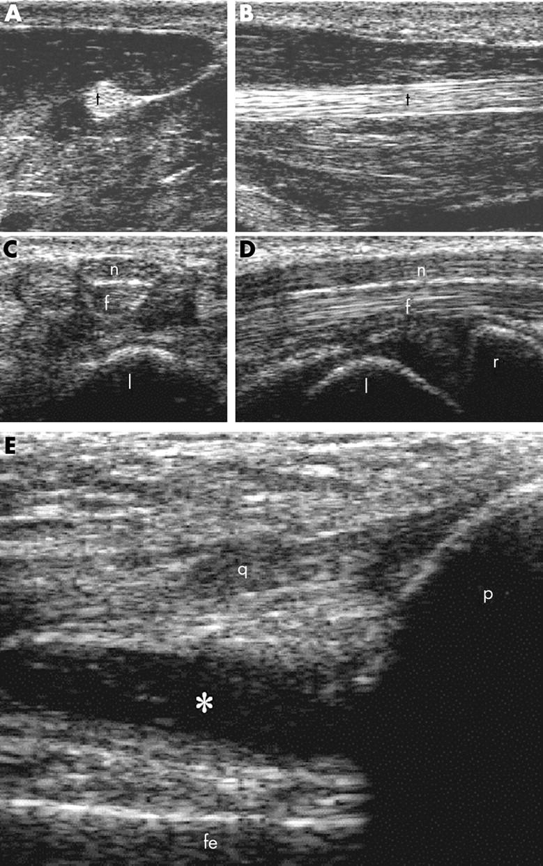
Figure 1.

(A, B) Healthy subject. Thenar eminence. Deep flexor tendon of the first finger (t). Transverse (A) and longitudinal (B) volar scans. (C, D) Healthy subject. Carpal tunnel. Median nerve (n) and finger flexor tendons (f). Transverse (C) and longitudinal (D) volar scans. l = lunate bone; r = radius. (E) Rheumatoid arthritis. Knee joint. Longitudinal suprapatellar scan. Suprapatellar pouch enlargement due to knee synovitis. * = synovial fluid; q = quadriceps tendon; p = patella; fe = femur.


