

Abstract
Methods: Synovial biopsy specimens were obtained by needle arthroscopy from the knees of 10 patients with either RA or ReA. RNA was isolated from the biopsy specimens and cDNA synthesised for analysis using a customised cDNA macroarray. Confirmatory analysis was performed using in situ hybridisation on a second set of synovial samples.
Results: Two unique transcripts (ReXS1 and fibronectin) were consistently more abundant in ReA and three homologous transcripts were more abundant in RA. The latter all mapped within long interspersed nucleotide elements (LINE-1), that form one of the families of repetitive sequences in the human genome.
Conclusions: The abundance of transcripts containing LINE-1 in the RA synovium may be an epiphenomenon or may have pathogenic significance. Further work is required to determine the identity of the full length transcript(s) before its use as a diagnostic marker in RA can be assessed.
Full Text
The Full Text of this article is available as a PDF (202.8 KB).
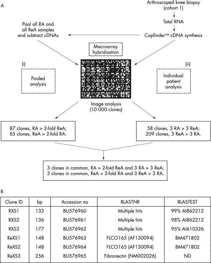
Figure 1. Summary of cDNA macroarray analysis. (A) A comparison of gene expression was carried out initially between (i) pooled, enriched cDNAs (two grids used), and (ii) cDNAs from patients RA1–3 and ReA1–3 (six grids used). The clones that gave consistent differences between RA and ReA samples and were common to both the pooled and individual patient analyses were selected. (B) Sequence analysis of the clones. The number of bases of cloned insert DNA is shown. The six clones (RXS1–3 and ReXS1–3 with accession numbers BU576960–5 respectively) were searched against the GenBank nucleotide database (BLASTNR) as well as the human EST (expressed sequence) database (BLASTEST). The accession numbers of the best matches are depicted. ND depicts not done.
Figure 2.
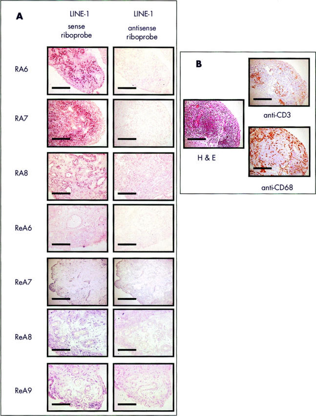
In situ hybridisation and immunohistology. (A) RXS1 in situ hybridisation. Serial sections of inflamed synovium from patients RA6–RA8 and ReA6–ReA9 were analysed using LINE-1 sense and antisense riboprobes. The images depicted are x200 magnification and the bar marker represents 50 µm. Note that RA6 and RA7 gave intense sublining layer staining for the LINE-1 sense riboprobe. (B) Patient RA7 immunohistology. Serial sections were analysed for general histology (haematoxylin and eosin), T cells (anti-CD3), and macrophages (anti-CD68), and the staining compared with the in situ hybridisation pattern. Standard histology depicts a lymphoid follicle and coincides with surrounding cells that give predominant in situ hybridisation staining with the LINE-1 sense riboprobe.