Abstract
Methods: 27 patients with SSc without clinical evidence of ischaemic heart disease and 23 control group subjects matched for age and sex were studied. CFR was evaluated in the left anterior descending coronary artery (LAD) with a new non-invasive method: contrast (Levovist) enhanced transthoracic Doppler during adenosine infusion. The pulsed wave Doppler examination of blood flow velocity was recorded in the LAD at rest and after maximum vasodilatation by adenosine infusion.
Results: In patients with SSc, without clinical evidence of ischaemic heart disease, CFR was impaired (p=0.0001). 14/27 patients with SSc had severe reduction of the CFR (⩽2.5) compared with controls (p=0.002). A non-significant trend between mean CFR and the severity and duration of the disease was also seen.
Conclusions: CFR is often reduced in patients with SSc, suggesting early preclinical cardiac involvement in SSc. This impairment in coronary microvasculature is detectable by a non-invasive echocardiographic method and in this study was more common in the diffuse form of SSc.
Full Text
The Full Text of this article is available as a PDF (114.1 KB).
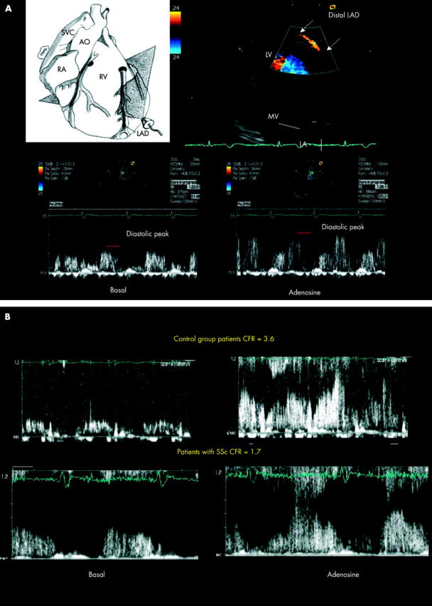
Figure 1.

(A) In the upper part of the figure a tomographic plane of a modified two chamber view is shown (at the left). Colour Doppler flow mapping in the distal portion of the LAD during contrast enhancement is shown (at the right). In the lower part, spectral Doppler tracing by sampling in the distal LAD at baseline and after adenosine intravenous injection is seen. A biphasic flow with a prevalent diastolic component is clearly detectable. LAD, left anterior descending coronary artery; LV, left ventricle; MV, mitral valve; LA, left atrium; SVC, superior vena cava; RA, right atrium; AO, aorta; RV, right ventricle; CFR, coronary flow reserve. (B) An example of a control group patient with normal CFR (upper section) and a sclerodermic patient with abnormal CFR (lower section). Enhanced transthoracic spectral Doppler flow at baseline (left) and during hyperaemia (right) are seen in both sections.
Figure 2.

Histogram illustrating CFR (mean of values) in control group and in patients with systemic sclerosis (upper panel). Histogram illustrating the percentage of CFR <2.5 in the control group and in patients with systemic sclerosis (lower panel).


