Abstract
Methods: Demographic, clinical data, and imaging data on hand radiographs and Genants vertebral deformity score on spine radiographs were collected from 135 women with RA of ⩾5 years, recruited from three European rheumatology clinics. Metacarpal hand BMD was measured by digital hand x ray radiogrammetry (DXR), and hip and lumbar spine BMD by dual x ray absorptiometry (DXA). Multiple regression analyses were used to examine associations between hand BMD and radiographic joint damage, and hand BMD and fractures.
Results: Hand BMD was strongly and independently associated with radiographic hand joint damage in a linear regression model adjusted for age, centre, BMI, disease duration, RF, 18 deformed joint count, ESR, and femoral neck BMD. In a multivariate logistic regression model adjusted for relevant variables, hand BMD and femoral neck BMD, but not spine BMD, were independently associated with vertebral deformities and with non-vertebral fractures.
Conclusion: BMD measured by DXR on conventional hand radiographs in patients with RA may potentially be used as an indicator of joint damage and of vertebral and non-vertebral fracture risk.
Full Text
The Full Text of this article is available as a PDF (91.9 KB).
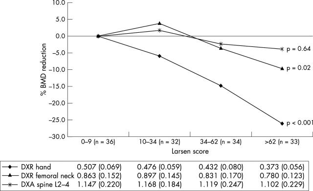
Figure 1.
Mean percentage reduction of BMD at hands, femoral neck, and spine (L2–4) related to quartile groups of radiographic damage using the lowest quartile of Larsen score as reference category. Below the figure mean (SD) BMD (g/cm2) values for each quartile of the Larsen score are shown. p Values from overall ANOVA (statistically significant group differences using post hoc Bonferroni test): hand BMD: 1st quartile v 3rd and 4th quartile, 2nd quartile v 3rd and 4th quartile, and 3rd quartile v 1st, 2nd, and 4th quartile; femoral neck BMD: 2nd quartile v 4th quartile.


