Abstract
Objective: To study the correlation between ankle and knee cartilage morphology to test the hypothesis that knee joint cartilage loss in gonarthritis can be estimated retrospectively using quantitative MRI analysis of the knee and ankle and established regression equations; and to test the hypothesis that sex differences in joint surface area are larger in the knee than the ankle, which may explain the greater incidence of knee osteoarthritis in elderly women than in elderly men.
Methods: Sagittal MR images (3D FLASH WE) of the knee and hind foot were acquired in 29 healthy subjects (14 women, 15 men; mean (SD) age, 25 (3) years), with no signs joint disease. Cartilage volume, thickness, and joint surface area were determined in the knee, ankle, and subtalar joint.
Results: Knee cartilage volumes and joint surface areas showed only moderate correlations with those of the ankle and subtalar joint (r = 0.33 to 0.81). The correlations of cartilage thickness between the two joints were weaker still (r = –0.05 to 0.53). Sex differences in cartilage morphology at the knee and the ankle were similar, with surface areas being –17.5% to –23.5% lower in women than in men.
Conclusions: Only moderate correlations in cartilage morphology of healthy subjects were found between knee and ankle. It is therefore impractical to estimate knee joint cartilage loss a posteriori in cross sectional studies by measuring the hind foot and then applying a scaling factor. Sex differences in cartilage morphology do not explain differences in osteoarthritis incidence between men and women in the knee and ankle.
Full Text
The Full Text of this article is available as a PDF (214.8 KB).
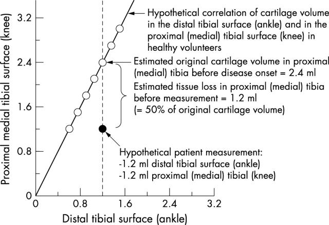
Figure 1.
Scattergram showing the rationale for the study. If the hypothesis is correct that there is a strong linear relation between knee joint cartilage volume in the knee and ankle surfaces (for example, medial tibia and distal tibia) in healthy volunteers (empty circles), this relation can be exploited to estimate retrospectively the amount of tissue loss in the knee surface in a cross sectional study. In the 29 healthy volunteers examined in this study, the cartilage volume of the medial tibia (knee joint) varied from 1.1 to 3.2 ml (mean 2.3 ml), and that of the distal tibia (ankle joint) from 0.6 to 1.7 ml (mean 1.1 ml). The graph also shows the hypothetical measurement in a patient (filled circle). Assuming a strong linear relation in healthy volunteers (empty circles), one could accurately estimate the cartilage tissue loss in the medial tibia of the hypothetical patient to amount to 1.2 ml (50% cartilage loss), although the measurement at the knee of this patients falls within the normal range.
Figure 2.

Sagittal magnetic resonance images of the knee and ankle, acquired with a high resolution FLASH (fast low angle shot) wider excitation sequence. The figure also displays the surfaces that have been analysed in the current study.
Figure 3.

Bivariate scattergrams showing the correlation of cartilage volume in the knee, ankle, and subtalar joint. Women and men are identified in the graphs. Surface, joint surface area; thickness, mean cartilage thickness; volume, cartilage volume.
Selected References
These references are in PubMed. This may not be the complete list of references from this article.
- Al-Ali Dina, Graichen Heiko, Faber Sonja, Englmeier Karl-Hans, Reiser Maximilian, Eckstein Felix. Quantitative cartilage imaging of the human hind foot: precision and inter-subject variability. J Orthop Res. 2002 Mar;20(2):249–256. doi: 10.1016/S0736-0266(01)00098-5. [DOI] [PubMed] [Google Scholar]
- Burgkart R., Glaser C., Hinterwimmer S., Hudelmaier M., Englmeier K-H, Reiser M., Eckstein F. Feasibility of T and Z scores from magnetic resonance imaging data for quantification of cartilage loss in osteoarthritis. Arthritis Rheum. 2003 Oct;48(10):2829–2835. doi: 10.1002/art.11259. [DOI] [PubMed] [Google Scholar]
- Burgkart R., Glaser C., Hyhlik-Dürr A., Englmeier K. H., Reiser M., Eckstein F. Magnetic resonance imaging-based assessment of cartilage loss in severe osteoarthritis: accuracy, precision, and diagnostic value. Arthritis Rheum. 2001 Sep;44(9):2072–2077. doi: 10.1002/1529-0131(200109)44:9<2072::AID-ART357>3.0.CO;2-3. [DOI] [PubMed] [Google Scholar]
- Cicuttini F. M., Wluka A. E., Forbes A., Wolfe R. Comparison of tibial cartilage volume and radiologic grade of the tibiofemoral joint. Arthritis Rheum. 2003 Mar;48(3):682–688. doi: 10.1002/art.10840. [DOI] [PubMed] [Google Scholar]
- Cicuttini Flavia, Wluka Anita, Wang Yuanyuan, Stuckey Stephen. The determinants of change in patella cartilage volume in osteoarthritic knees. J Rheumatol. 2002 Dec;29(12):2615–2619. [PubMed] [Google Scholar]
- Eckstein F., Gavazzeni A., Sittek H., Haubner M., Lösch A., Milz S., Englmeier K. H., Schulte E., Putz R., Reiser M. Determination of knee joint cartilage thickness using three-dimensional magnetic resonance chondro-crassometry (3D MR-CCM). Magn Reson Med. 1996 Aug;36(2):256–265. doi: 10.1002/mrm.1910360213. [DOI] [PubMed] [Google Scholar]
- Eckstein F., Heudorfer L., Faber S. C., Burgkart R., Englmeier K-H, Reiser M. Long-term and resegmentation precision of quantitative cartilage MR imaging (qMRI). Osteoarthritis Cartilage. 2002 Dec;10(12):922–928. doi: 10.1053/joca.2002.0844. [DOI] [PubMed] [Google Scholar]
- Eckstein F., Müller S., Faber S. C., Englmeier K-H, Reiser M., Putz R. Side differences of knee joint cartilage volume, thickness, and surface area, and correlation with lower limb dominance--an MRI-based study. Osteoarthritis Cartilage. 2002 Dec;10(12):914–921. doi: 10.1053/joca.2002.0843. [DOI] [PubMed] [Google Scholar]
- Eckstein F., Winzheimer M., Hohe J., Englmeier K. H., Reiser M. Interindividual variability and correlation among morphological parameters of knee joint cartilage plates: analysis with three-dimensional MR imaging. Osteoarthritis Cartilage. 2001 Feb;9(2):101–111. doi: 10.1053/joca.2000.0365. [DOI] [PubMed] [Google Scholar]
- Eger Wolfgang, Schumacher Barbara L., Mollenhauer Jürgen, Kuettner Klaus E., Cole Ada A. Human knee and ankle cartilage explants: catabolic differences. J Orthop Res. 2002 May;20(3):526–534. doi: 10.1016/S0736-0266(01)00125-5. [DOI] [PubMed] [Google Scholar]
- Faber S. C., Eckstein F., Lukasz S., Mühlbauer R., Hohe J., Englmeier K. H., Reiser M. Gender differences in knee joint cartilage thickness, volume and articular surface areas: assessment with quantitative three-dimensional MR imaging. Skeletal Radiol. 2001 Mar;30(3):144–150. doi: 10.1007/s002560000320. [DOI] [PubMed] [Google Scholar]
- Felson D. T., Zhang Y., Hannan M. T., Naimark A., Weissman B. N., Aliabadi P., Levy D. The incidence and natural history of knee osteoarthritis in the elderly. The Framingham Osteoarthritis Study. Arthritis Rheum. 1995 Oct;38(10):1500–1505. doi: 10.1002/art.1780381017. [DOI] [PubMed] [Google Scholar]
- Gandy S. J., Dieppe P. A., Keen M. C., Maciewicz R. A., Watt I., Waterton J. C. No loss of cartilage volume over three years in patients with knee osteoarthritis as assessed by magnetic resonance imaging. Osteoarthritis Cartilage. 2002 Dec;10(12):929–937. doi: 10.1053/joca.2002.0849. [DOI] [PubMed] [Google Scholar]
- Glaser C., Faber S., Eckstein F., Fischer H., Springer V., Heudorfer L., Stammberger T., Englmeier K. H., Reiser M. Optimization and validation of a rapid high-resolution T1-w 3D FLASH water excitation MRI sequence for the quantitative assessment of articular cartilage volume and thickness. Magn Reson Imaging. 2001 Feb;19(2):177–185. doi: 10.1016/s0730-725x(01)00292-2. [DOI] [PubMed] [Google Scholar]
- Graichen H., Jakob J., von Eisenhart-Rothe R., Englmeier K-H, Reiser M., Eckstein F. Validation of cartilage volume and thickness measurements in the human shoulder with quantitative magnetic resonance imaging. Osteoarthritis Cartilage. 2003 Jul;11(7):475–482. doi: 10.1016/s1063-4584(03)00077-3. [DOI] [PubMed] [Google Scholar]
- Graichen H., Springer V., Flaman T., Stammberger T., Glaser C., Englmeier K. H., Reiser M., Eckstein F. Validation of high-resolution water-excitation magnetic resonance imaging for quantitative assessment of thin cartilage layers. Osteoarthritis Cartilage. 2000 Mar;8(2):106–114. doi: 10.1053/joca.1999.0278. [DOI] [PubMed] [Google Scholar]
- Graichen Heiko, von Eisenhart-Rothe Rüdiger, Vogl Thomas, Englmeier Karl-Hans, Eckstein Felix. Quantitative assessment of cartilage status in osteoarthritis by quantitative magnetic resonance imaging: technical validation for use in analysis of cartilage volume and further morphologic parameters. Arthritis Rheum. 2004 Mar;50(3):811–816. doi: 10.1002/art.20191. [DOI] [PubMed] [Google Scholar]
- Hohe Jan, Ateshian Gerard, Reiser Maximilian, Englmeier Karl-Hans, Eckstein Felix. Surface size, curvature analysis, and assessment of knee joint incongruity with MRI in vivo. Magn Reson Med. 2002 Mar;47(3):554–561. doi: 10.1002/mrm.10097. [DOI] [PubMed] [Google Scholar]
- Hohe Jan, Faber Sonja, Muehlbauer Roland, Reiser Maximilian, Englmeier Karl-Hans, Eckstein Felix. Three-dimensional analysis and visualization of regional MR signal intensity distribution of articular cartilage. Med Eng Phys. 2002 Apr;24(3):219–227. doi: 10.1016/s1350-4533(02)00006-1. [DOI] [PubMed] [Google Scholar]
- Huch K. Knee and ankle: human joints with different susceptibility to osteoarthritis reveal different cartilage cellularity and matrix synthesis in vitro. Arch Orthop Trauma Surg. 2001 Jun;121(6):301–306. doi: 10.1007/s004020000225. [DOI] [PubMed] [Google Scholar]
- Huch K., Kuettner K. E., Dieppe P. Osteoarthritis in ankle and knee joints. Semin Arthritis Rheum. 1997 Feb;26(4):667–674. doi: 10.1016/s0049-0172(97)80002-9. [DOI] [PubMed] [Google Scholar]
- Hudelmaier M., Glaser C., Hohe J., Englmeier K. H., Reiser M., Putz R., Eckstein F. Age-related changes in the morphology and deformational behavior of knee joint cartilage. Arthritis Rheum. 2001 Nov;44(11):2556–2561. doi: 10.1002/1529-0131(200111)44:11<2556::aid-art436>3.0.co;2-u. [DOI] [PubMed] [Google Scholar]
- Hudelmaier Martin, Glaser Christian, Englmeier Karl-Hans, Reiser Maximilian, Putz Reinhard, Eckstein Felix. Correlation of knee-joint cartilage morphology with muscle cross-sectional areas vs. anthropometric variables. Anat Rec A Discov Mol Cell Evol Biol. 2003 Feb;270(2):175–184. doi: 10.1002/ar.a.10001. [DOI] [PubMed] [Google Scholar]
- Jones G., Glisson M., Hynes K., Cicuttini F. Sex and site differences in cartilage development: a possible explanation for variations in knee osteoarthritis in later life. Arthritis Rheum. 2000 Nov;43(11):2543–2549. doi: 10.1002/1529-0131(200011)43:11<2543::AID-ANR23>3.0.CO;2-K. [DOI] [PubMed] [Google Scholar]
- Jones Graeme, Ding Changhai, Glisson Michael, Hynes Kristen, Ma Deqiong, Cicuttini Flavia. Knee articular cartilage development in children: a longitudinal study of the effect of sex, growth, body composition, and physical activity. Pediatr Res. 2003 May 7;54(2):230–236. doi: 10.1203/01.PDR.0000072781.93856.E6. [DOI] [PubMed] [Google Scholar]
- Kang Y., Koepp H., Cole A. A., Kuettner K. E., Homandberg G. A. Cultured human ankle and knee cartilage differ in susceptibility to damage mediated by fibronectin fragments. J Orthop Res. 1998 Sep;16(5):551–556. doi: 10.1002/jor.1100160505. [DOI] [PubMed] [Google Scholar]
- Kerin A., Patwari P., Kuettner K., Cole A., Grodzinsky A. Molecular basis of osteoarthritis: biomechanical aspects. Cell Mol Life Sci. 2002 Jan;59(1):27–35. doi: 10.1007/s00018-002-8402-1. [DOI] [PMC free article] [PubMed] [Google Scholar]
- Koepp H., Eger W., Muehleman C., Valdellon A., Buckwalter J. A., Kuettner K. E., Cole A. A. Prevalence of articular cartilage degeneration in the ankle and knee joints of human organ donors. J Orthop Sci. 1999;4(6):407–412. doi: 10.1007/s007760050123. [DOI] [PubMed] [Google Scholar]
- Nevitt M. C., Felson D. T., Williams E. N., Grady D. The effect of estrogen plus progestin on knee symptoms and related disability in postmenopausal women: The Heart and Estrogen/Progestin Replacement Study, a randomized, double-blind, placebo-controlled trial. Arthritis Rheum. 2001 Apr;44(4):811–818. doi: 10.1002/1529-0131(200104)44:4<811::AID-ANR137>3.0.CO;2-F. [DOI] [PubMed] [Google Scholar]
- Peterfy Charles G. Imaging of the disease process. Curr Opin Rheumatol. 2002 Sep;14(5):590–596. doi: 10.1097/00002281-200209000-00020. [DOI] [PubMed] [Google Scholar]
- Stammberger T., Eckstein F., Englmeier K. H., Reiser M. Determination of 3D cartilage thickness data from MR imaging: computational method and reproducibility in the living. Magn Reson Med. 1999 Mar;41(3):529–536. doi: 10.1002/(sici)1522-2594(199903)41:3<529::aid-mrm15>3.0.co;2-z. [DOI] [PubMed] [Google Scholar]
- Stammberger T., Eckstein F., Michaelis M., Englmeier K. H., Reiser M. Interobserver reproducibility of quantitative cartilage measurements: comparison of B-spline snakes and manual segmentation. Magn Reson Imaging. 1999 Sep;17(7):1033–1042. doi: 10.1016/s0730-725x(99)00040-5. [DOI] [PubMed] [Google Scholar]
- Treppo S., Koepp H., Quan E. C., Cole A. A., Kuettner K. E., Grodzinsky A. J. Comparison of biomechanical and biochemical properties of cartilage from human knee and ankle pairs. J Orthop Res. 2000 Sep;18(5):739–748. doi: 10.1002/jor.1100180510. [DOI] [PubMed] [Google Scholar]
- Von Mühlen Denise, Morton Deborah, Von Mühlen Carlos A., Barrett-Connor Elizabeth. Postmenopausal estrogen and increased risk of clinical osteoarthritis at the hip, hand, and knee in older women. J Womens Health Gend Based Med. 2002 Jul-Aug;11(6):511–518. doi: 10.1089/152460902760277868. [DOI] [PubMed] [Google Scholar]
- Wluka Anita E., Stuckey Stephen, Snaddon Judith, Cicuttini Flavia M. The determinants of change in tibial cartilage volume in osteoarthritic knees. Arthritis Rheum. 2002 Aug;46(8):2065–2072. doi: 10.1002/art.10460. [DOI] [PubMed] [Google Scholar]


