Full Text
The Full Text of this article is available as a PDF (128.8 KB).
Figure 1 .
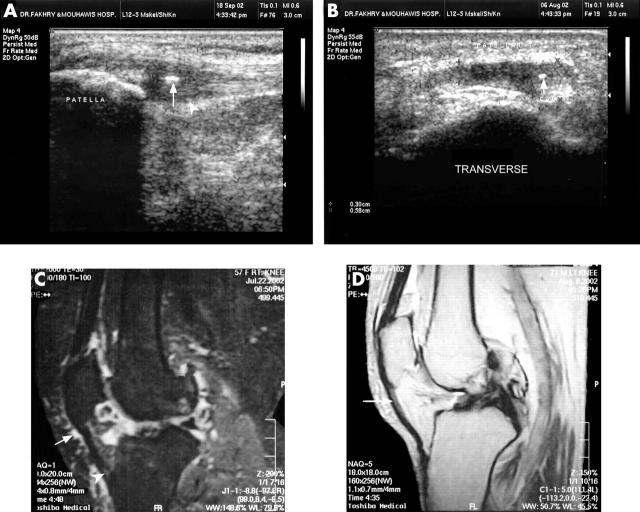
(A) A sagittal US scan shows thickened proximal entheses of the patellar ligament with loss of its fibrillar echo pattern, loss of the sharp definition of its posterior aspect compared with the distal portion (arrow head), calcific foci (arrow). (B) A transverse US scan of the same patient shows the thickened medial part of the patellar ligament with calcific focus (arrow). (C) A sagittal T2 fat suppression image shows the thickened distal part of the patellar tendon with altered signal intensity (arrow head) and prepatellar bursitis (arrow). (D) A sagittal Pd weighted image shows high intensity signals of the proximal patellar tendon.


