Abstract
Background: Bone mineral density (BMD) measurements are frequently performed repeatedly for each patient. Subsequent BMD measurements allow reproducibility to be assessed.
Objective: To examine the reproducibility of BMD by dual energy x ray absorptiometry (DXA) and to investigate the practical value of different measures of reproducibility in a group of postmenopausal women.
Methods: Ninety five women, mean age 59.9 years, underwent two subsequent BMD measurements of spine and hip. Reproducibility was expressed as smallest detectable difference (SDD), coefficient of variation (CV), and intraclass correlation coefficient (ICC). Sources of variation were investigated by multilevel analysis.
Results: The median interval between measurements was 0 days (range 0–45). The mean difference (SD) between the measurements (g/cm2) was -0.001 (0.02) and -0.0004 (0.02) at L1-4 and the total hip, respectively. At L1-4 and the total hip, SDD (g/cm2) was ±0.05 and ±0.04 and CV (%) was 1.92 and 1.59, respectively. The ICC at spine and hip was 0.99.
Conclusions: Reproducibility in the postmenopausal women studied was good. In a repeated DXA scan a BMD change exceeding 2√2CV (%), the least significant change (LSC), or the SDD should be regarded as significant. Use of the SDD is preferable to use of the CV and LSC (%) because of its independence from BMD and its expression in absolute units. Expressed as SDD, a BMD change of at least ±0.05 g/cm2 at L1-4 and ±0.04 g/cm2 at the total hip should be considered significant.
Full Text
The Full Text of this article is available as a PDF (291.3 KB).
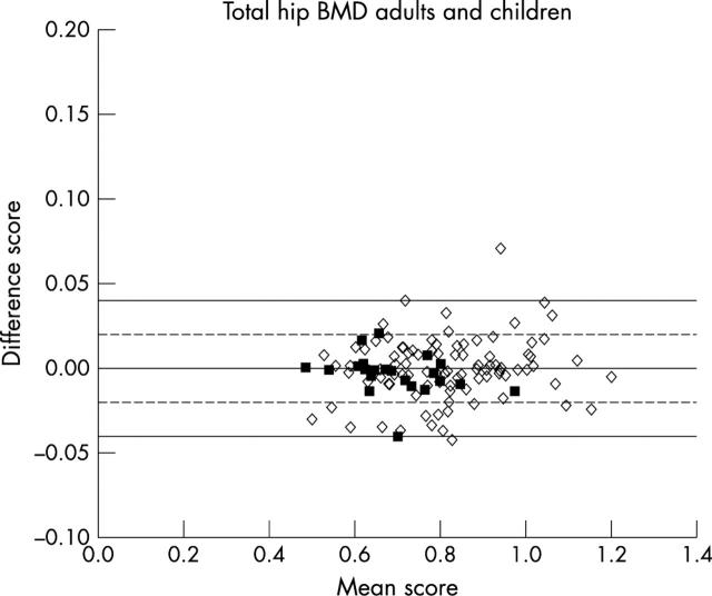
Figure 1 .
Graph of the difference score against the mean score of the two total hip BMD measurements (g/cm2) in postmenopausal women (open diamonds) and children (closed squares). The outermost (solid) lines represent the 95% limits of agreement for postmenopausal women. The inner (dashed) lines represent the 95% limits of agreement for children.
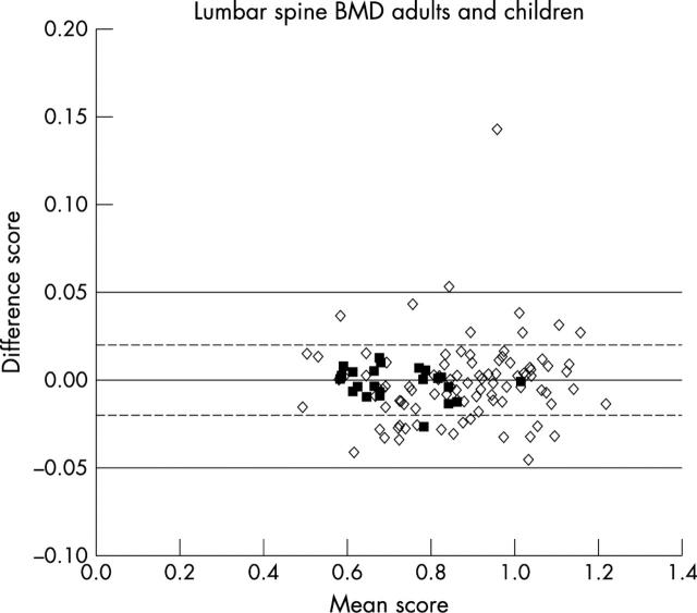
Figure 2 .
Graph of the difference score against the mean score of the two lumbar spine BMD measurements (g/cm2) in postmenopausal women (open diamonds) and children (closed squares). The outermost (solid) lines represent the 95% limits of agreement for postmenopausal women. The inner (dashed) lines represent the 95% limits of agreement for children.
Selected References
These references are in PubMed. This may not be the complete list of references from this article.
- Blake G. M., Fogelman I. Bone densitometry and the diagnosis of osteoporosis. Semin Nucl Med. 2001 Jan;31(1):69–81. doi: 10.1053/snuc.2001.18749. [DOI] [PubMed] [Google Scholar]
- Bland J. M., Altman D. G. Statistical methods for assessing agreement between two methods of clinical measurement. Lancet. 1986 Feb 8;1(8476):307–310. [PubMed] [Google Scholar]
- Boutsen Y., Jamart J., Esselinckx W., Devogelaer J. P. Primary prevention of glucocorticoid-induced osteoporosis with intravenous pamidronate and calcium: a prospective controlled 1-year study comparing a single infusion, an infusion given once every 3 months, and calcium alone. J Bone Miner Res. 2001 Jan;16(1):104–112. doi: 10.1359/jbmr.2001.16.1.104. [DOI] [PubMed] [Google Scholar]
- Cohen S., Levy R. M., Keller M., Boling E., Emkey R. D., Greenwald M., Zizic T. M., Wallach S., Sewell K. L., Lukert B. P. Risedronate therapy prevents corticosteroid-induced bone loss: a twelve-month, multicenter, randomized, double-blind, placebo-controlled, parallel-group study. Arthritis Rheum. 1999 Nov;42(11):2309–2318. doi: 10.1002/1529-0131(199911)42:11<2309::AID-ANR8>3.0.CO;2-K. [DOI] [PubMed] [Google Scholar]
- Cummings S. R., Black D. Should perimenopausal women be screened for osteoporosis? Ann Intern Med. 1986 Jun;104(6):817–823. doi: 10.7326/0003-4819-104-6-817. [DOI] [PubMed] [Google Scholar]
- Diamond T., McGuigan L., Barbagallo S., Bryant C. Cyclical etidronate plus ergocalciferol prevents glucocorticoid-induced bone loss in postmenopausal women. Am J Med. 1995 May;98(5):459–463. doi: 10.1016/S0002-9343(99)80345-3. [DOI] [PubMed] [Google Scholar]
- Eastell R. Assessment of bone density and bone loss. Osteoporos Int. 1996;6 (Suppl 2):3–5. doi: 10.1007/BF01625232. [DOI] [PubMed] [Google Scholar]
- Fuleihan G. E., Testa M. A., Angell J. E., Porrino N., Leboff M. S. Reproducibility of DXA absorptiometry: a model for bone loss estimates. J Bone Miner Res. 1995 Jul;10(7):1004–1014. doi: 10.1002/jbmr.5650100704. [DOI] [PubMed] [Google Scholar]
- Genant H. K., Block J. E., Steiger P., Glueer C. C., Ettinger B., Harris S. T. Appropriate use of bone densitometry. Radiology. 1989 Mar;170(3 Pt 1):817–822. doi: 10.1148/radiology.170.3.2916037. [DOI] [PubMed] [Google Scholar]
- Genant H. K., Cooper C., Poor G., Reid I., Ehrlich G., Kanis J., Nordin B. E., Barrett-Connor E., Black D., Bonjour J. P. Interim report and recommendations of the World Health Organization Task-Force for Osteoporosis. Osteoporos Int. 1999;10(4):259–264. doi: 10.1007/s001980050224. [DOI] [PubMed] [Google Scholar]
- Genant H. K., Engelke K., Fuerst T., Glüer C. C., Grampp S., Harris S. T., Jergas M., Lang T., Lu Y., Majumdar S. Noninvasive assessment of bone mineral and structure: state of the art. J Bone Miner Res. 1996 Jun;11(6):707–730. doi: 10.1002/jbmr.5650110602. [DOI] [PubMed] [Google Scholar]
- Glüer C. C., Blake G., Lu Y., Blunt B. A., Jergas M., Genant H. K. Accurate assessment of precision errors: how to measure the reproducibility of bone densitometry techniques. Osteoporos Int. 1995;5(4):262–270. doi: 10.1007/BF01774016. [DOI] [PubMed] [Google Scholar]
- Glüer C. C. Monitoring skeletal changes by radiological techniques. J Bone Miner Res. 1999 Nov;14(11):1952–1962. doi: 10.1359/jbmr.1999.14.11.1952. [DOI] [PubMed] [Google Scholar]
- Glüer C. C., Steiger P., Selvidge R., Elliesen-Kliefoth K., Hayashi C., Genant H. K. Comparative assessment of dual-photon absorptiometry and dual-energy radiography. Radiology. 1990 Jan;174(1):223–228. doi: 10.1148/radiology.174.1.2294552. [DOI] [PubMed] [Google Scholar]
- Kanis J. A., Melton L. J., 3rd, Christiansen C., Johnston C. C., Khaltaev N. The diagnosis of osteoporosis. J Bone Miner Res. 1994 Aug;9(8):1137–1141. doi: 10.1002/jbmr.5650090802. [DOI] [PubMed] [Google Scholar]
- Kelly T. L., Slovik D. M., Schoenfeld D. A., Neer R. M. Quantitative digital radiography versus dual photon absorptiometry of the lumbar spine. J Clin Endocrinol Metab. 1988 Oct;67(4):839–844. doi: 10.1210/jcem-67-4-839. [DOI] [PubMed] [Google Scholar]
- Lees B., Stevenson J. C. An evaluation of dual-energy X-ray absorptiometry and comparison with dual-photon absorptiometry. Osteoporos Int. 1992 May;2(3):146–152. doi: 10.1007/BF01623822. [DOI] [PubMed] [Google Scholar]
- Maggio D., McCloskey E. V., Camilli L., Cenci S., Cherubini A., Kanis J. A., Senin U. Short-term reproducibility of proximal femur bone mineral density in the elderly. Calcif Tissue Int. 1998 Oct;63(4):296–299. doi: 10.1007/s002239900530. [DOI] [PubMed] [Google Scholar]
- Miller P. D., Bonnick S. L., Rosen C. J. Consensus of an international panel on the clinical utility of bone mass measurements in the detection of low bone mass in the adult population. Calcif Tissue Int. 1996 Apr;58(4):207–214. doi: 10.1007/BF02508636. [DOI] [PubMed] [Google Scholar]
- Neele S. J., Marchien van Baal W., van der Mooren M. J., Kessel H., Netelenbos J. C., Kenemans P. Ultrasound assessment of the endometrium in healthy, asymptomatic early post-menopausal women: saline infusion sonohysterography versus transvaginal ultrasound. Ultrasound Obstet Gynecol. 2000 Sep;16(3):254–259. doi: 10.1046/j.1469-0705.2000.00273.x. [DOI] [PubMed] [Google Scholar]
- Nguyen T. V., Eisman J. A. Assessment of significant change in BMD: a new approach. J Bone Miner Res. 2000 Feb;15(2):369–372. doi: 10.1359/jbmr.2000.15.2.369. [DOI] [PubMed] [Google Scholar]
- Oleksik A., Ott S. M., Vedi S., Bravenboer N., Compston J., Lips P. Bone structure in patients with low bone mineral density with or without vertebral fractures. J Bone Miner Res. 2000 Jul;15(7):1368–1375. doi: 10.1359/jbmr.2000.15.7.1368. [DOI] [PubMed] [Google Scholar]
- Pouilles J. M., Tremollieres F., Todorovsky N., Ribot C. Precision and sensitivity of dual-energy x-ray absorptiometry in spinal osteoporosis. J Bone Miner Res. 1991 Sep;6(9):997–1002. doi: 10.1002/jbmr.5650060914. [DOI] [PubMed] [Google Scholar]
- Ravaud P., Reny J. L., Giraudeau B., Porcher R., Dougados M., Roux C. Individual smallest detectable difference in bone mineral density measurements. J Bone Miner Res. 1999 Aug;14(8):1449–1456. doi: 10.1359/jbmr.1999.14.8.1449. [DOI] [PubMed] [Google Scholar]
- Slosman D. O., Rizzoli R., Bonjour J. P. Bone absorptiometry: a critical appraisal of various methods. Acta Paediatr Suppl. 1995 Sep;411:9–11. doi: 10.1111/j.1651-2227.1995.tb13851.x. [DOI] [PubMed] [Google Scholar]
- Van Coeverden S. C., De Ridder C. M., Roos J. C., Van't Hof M. A., Netelenbos J. C., Delemarre-Van de Waal H. A. Pubertal maturation characteristics and the rate of bone mass development longitudinally toward menarche. J Bone Miner Res. 2001 Apr;16(4):774–781. doi: 10.1359/jbmr.2001.16.4.774. [DOI] [PubMed] [Google Scholar]
- de Valk-de Roo G. W., Stehouwer C. D., Lambert J., Schalkwijk C. G., van der Mooren M. J., Kluft C., Netelenbos C. Plasma homocysteine is weakly correlated with plasma endothelin and von Willebrand factor but not with endothelium-dependent vasodilatation in healthy postmenopausal women. Clin Chem. 1999 Aug;45(8 Pt 1):1200–1205. [PubMed] [Google Scholar]


