

Abstract
Materials and methods: Aging male DBA/1 mice from different litters were caged together (4–6 mice per cage) at the age of 12 weeks, checked twice a week for signs of arthritis, and killed at different times. Hind paws were dissected and processed for histology.
Results: Disease incidence varied between 50% and 100% in four different experiments. Besides clinical signs of arthritis, nail abnormalities were noticed. Pathological examination showed the occurrence of dactylitis characterised by diffuse neutrophil infiltration in 6 of 50 paws examined. Onycho-periostitis with progressive destruction of the nail bed and the underlying distal phalanx was seen in 5 of 50 paws examined.
Conclusions: Although dactylitis and onychoperiostitis are rare manifestations of the disease process, these data strongly suggest that spontaneous arthritis in aging male DBA/1 mice shares important features with human psoriatic arthritis. This model may therefore be an important tool to study links between stress, sex, inflammation, and new bone formation with particular relevance to human psoriatic arthritis.
Full Text
The Full Text of this article is available as a PDF (799.8 KB).
Figure 1 .
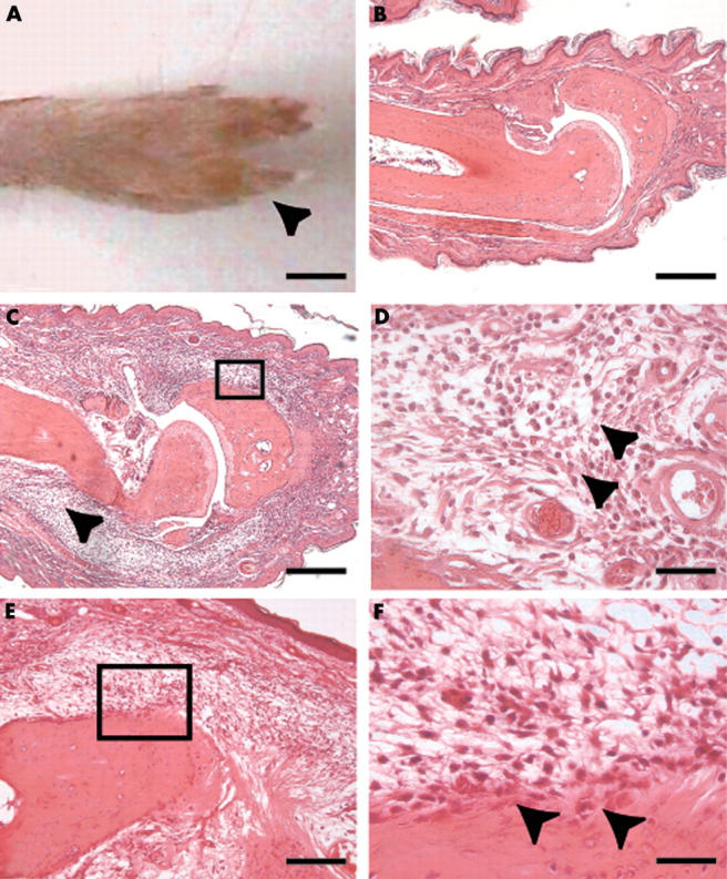
Dactylitis in spontaneous arthritis in aging male DBA/1 mice. (A) clinical signs of dactylitis (sausage digit) in the 4th toe (arrowhead); (B) microscopic image of a normal toe with a proximal interphalangeal joint; (C) microscopic image of dactylitis showing extensive subcutaneous oedema and tenosynovitis with disruption of muscle and tendon fibres (arrowhead); (D) microscopic detail showing neutrophils (arrowheads) in the subcutaneous tissue; (E) inflammatory reaction adjacent to the enthesis; (F) microscopic detail showing polynuclear and mononuclear cells (arrowheads) at the enthesis. (A) Bar = 2.5 mm. Haematoxylin and eosin staining; bar = 400 µm (B, C), 200 µm (E), and 50 µm (D-F).
Figure 2 .
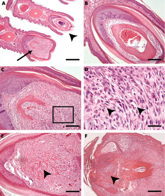
Onychoperiostitis in spontaneous arthritis in aging male DBA/1 mice. (A) Overview showing a normal toe and nail with the underlying distal phalanx (arrowhead) as well as a toe with an affected nail and distal phalanx periostitis (arrow); (B) detail of normal nail bed and distal phalanx ; (C and E) periostitis in diseased nails showing a hypercellular tissue (C) and bone destructive, pannus-like tissue (E) invading the underlying distal phalanx (arrowhead); (D) detail from C showing spindle shaped cells and some small blood vessels (arrowheads); (F) late stage onychoperiostitis resulting in complete destruction of the nail bed and the distal phalanx (arrowhead). Haematoxylin and eosin staining; bar = 800 µm (A), 400 µm (F), 200 µm (B, C, E), and 50 µm (D).