Abstract
Objectives: To determine whether magnetic resonance (MR) scans of the dominant wrist of patients with early rheumatoid arthritis (RA) can be used to predict functional outcome at 6 years' follow up.
Methods: Dominant wrist MR scans were obtained in 42 patients with criteria for RA at first presentation. Patients were followed up prospectively for 6 years, and further scans obtained at 1 year (42 patients) and 6 years (31 patients). Two radiologists scored scans for synovitis, tendonitis, bone oedema, and erosions. The Stanford Health Assessment Questionnaire (HAQ) score, indicating functional outcome, and standard measures of disease activity were assessed at 0, 1, 2, and 6 years. The physical function component of the SF-36 score (PF-SF36) was also used as a functional outcome measure at 6 years.
Results: Baseline MR parameters, including bone oedema score and the total baseline MR score, were predictive of the PF-SF36 at 6 years (R2 = 0.22, p = 0.005 and R2 = 0.16, p = 0.02, respectively). The PF-SF36 score correlated strongly with the HAQ score at 6 years (rs = –0.725, p<0.0001); none of the baseline MR parameters predicted the 6 year HAQ score. The total MR score obtained at 1 year was predictive of the 6 year HAQ (R2 = 0.04, p = 0.01). Standard clinical and radiographic measures at baseline were not predictive of the 6 year PF-SF36, but when combined in a model with baseline MR oedema score, prediction increased from 0.09 to 0.23, or 23% of the 6 year variance.
Conclusion: MR imaging of the wrist in patients with early RA can help to predict function at 6 years and could be used to plan aggressive management at an earlier stage.
Full Text
The Full Text of this article is available as a PDF (353.9 KB).
Figure 1 .

Progression of MR, clinical, and functional parameters over 6 years. Solid lines indicate median scores at 0, 1, and 6 years for MR parameters and 0, 1, 2, and 6 years for DAS and HAQ. Dotted lines indicate interquartile ranges.
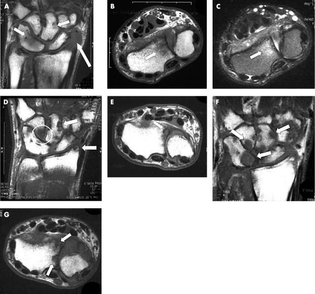
Figure 2 .
Sequential MR scans of the dominant carpus over 6 years in a 44 year old patient who was non-compliant with disease modifying treatment. This patient has congenital fusion of the triquetrum and lunate. (A) The baseline T1 weighted coronal scan shows focal areas of bone oedema as low signal within the capitate and the triquetrum/lunate (short arrows). There is extensive synovitis adjacent to the scaphoid (long arrow). (B) Axial T1 weighted image (baseline) showing bone oedema as low signal within the distal radius. (C) Equivalent baseline axial T2 weighted image showing bone oedema within the radius as bright signal. (D) Coronal T1 weighted scan at 1 year showing discrete low signal regions breaching the cortex of the capitate and the radial styloid indicating erosions (arrows). There is also extensive bone oedema within the hamate (circle). (E) Axial T1 weighted image at 1 year showing erosion at the radius. (F) Coronal T1 weighted scan at 6 years showing extensive erosive disease at multiple sites (arrows). (G) Axial T1 weighted image at 6 years showing more extensive erosion at the radius.
Selected References
These references are in PubMed. This may not be the complete list of references from this article.
- Arnett F. C., Edworthy S. M., Bloch D. A., McShane D. J., Fries J. F., Cooper N. S., Healey L. A., Kaplan S. R., Liang M. H., Luthra H. S. The American Rheumatism Association 1987 revised criteria for the classification of rheumatoid arthritis. Arthritis Rheum. 1988 Mar;31(3):315–324. doi: 10.1002/art.1780310302. [DOI] [PubMed] [Google Scholar]
- Boutry Nathalie, Lardé Anne, Lapègue Franck, Solau-Gervais Elizabeth, Flipo René-Marc, Cotten Anne. Magnetic resonance imaging appearance of the hands and feet in patients with early rheumatoid arthritis. J Rheumatol. 2003 Apr;30(4):671–679. [PubMed] [Google Scholar]
- Buchbinder R., Bombardier C., Yeung M., Tugwell P. Which outcome measures should be used in rheumatoid arthritis clinical trials? Clinical and quality-of-life measures' responsiveness to treatment in a randomized controlled trial. Arthritis Rheum. 1995 Nov;38(11):1568–1580. doi: 10.1002/art.1780381108. [DOI] [PubMed] [Google Scholar]
- Combe B., Eliaou J. F., Daurès J. P., Meyer O., Clot J., Sany J. Prognostic factors in rheumatoid arthritis. Comparative study of two subsets of patients according to severity of articular damage. Br J Rheumatol. 1995 Jun;34(6):529–534. doi: 10.1093/rheumatology/34.6.529. [DOI] [PubMed] [Google Scholar]
- Drossaers-Bakker K. W., de Buck M., van Zeben D., Zwinderman A. H., Breedveld F. C., Hazes J. M. Long-term course and outcome of functional capacity in rheumatoid arthritis: the effect of disease activity and radiologic damage over time. Arthritis Rheum. 1999 Sep;42(9):1854–1860. doi: 10.1002/1529-0131(199909)42:9<1854::AID-ANR9>3.0.CO;2-F. [DOI] [PubMed] [Google Scholar]
- Feigenbaum S. L., Masi A. T., Kaplan S. B. Prognosis in rheumatoid arthritis. A longitudinal study of newly diagnosed younger adult patients. Am J Med. 1979 Mar;66(3):377–384. doi: 10.1016/0002-9343(79)91055-6. [DOI] [PubMed] [Google Scholar]
- Foley-Nolan D., Stack J. P., Ryan M., Redmond U., Barry C., Ennis J., Coughlan R. J. Magnetic resonance imaging in the assessment of rheumatoid arthritis--a comparison with plain film radiographs. Br J Rheumatol. 1991 Apr;30(2):101–106. doi: 10.1093/rheumatology/30.2.101. [DOI] [PubMed] [Google Scholar]
- Fries J. F., Spitz P., Kraines R. G., Holman H. R. Measurement of patient outcome in arthritis. Arthritis Rheum. 1980 Feb;23(2):137–145. doi: 10.1002/art.1780230202. [DOI] [PubMed] [Google Scholar]
- Fuchs H. A., Kaye J. J., Callahan L. F., Nance E. P., Pincus T. Evidence of significant radiographic damage in rheumatoid arthritis within the first 2 years of disease. J Rheumatol. 1989 May;16(5):585–591. [PubMed] [Google Scholar]
- Gilkeson G., Polisson R., Sinclair H., Vogler J., Rice J., Caldwell D., Spritzer C., Martinez S. Early detection of carpal erosions in patients with rheumatoid arthritis: a pilot study of magnetic resonance imaging. J Rheumatol. 1988 Sep;15(9):1361–1366. [PubMed] [Google Scholar]
- Li G., Haq W., Xiang L., Lou B. S., Hughes R., De Leon I. A., Davis P., Gillespie T. J., Romanowski M., Zhu X. Modifications of the 4,4'-residues and SAR studies of Biphalin, a highly potent opioid receptor active peptide. Bioorg Med Chem Lett. 1998 Mar 3;8(5):555–560. doi: 10.1016/s0960-894x(98)00065-1. [DOI] [PubMed] [Google Scholar]
- McHorney C. A., Ware J. E., Jr, Lu J. F., Sherbourne C. D. The MOS 36-item Short-Form Health Survey (SF-36): III. Tests of data quality, scaling assumptions, and reliability across diverse patient groups. Med Care. 1994 Jan;32(1):40–66. doi: 10.1097/00005650-199401000-00004. [DOI] [PubMed] [Google Scholar]
- McQueen F. M., Benton N., Crabbe J., Robinson E., Yeoman S., McLean L., Stewart N. What is the fate of erosions in early rheumatoid arthritis? Tracking individual lesions using x rays and magnetic resonance imaging over the first two years of disease. Ann Rheum Dis. 2001 Sep;60(9):859–868. [PMC free article] [PubMed] [Google Scholar]
- McQueen F. M., Stewart N., Crabbe J., Robinson E., Yeoman S., Tan P. L., McLean L. Magnetic resonance imaging of the wrist in early rheumatoid arthritis reveals a high prevalence of erosions at four months after symptom onset. Ann Rheum Dis. 1998 Jun;57(6):350–356. doi: 10.1136/ard.57.6.350. [DOI] [PMC free article] [PubMed] [Google Scholar]
- McQueen F. M., Stewart N., Crabbe J., Robinson E., Yeoman S., Tan P. L., McLean L. Magnetic resonance imaging of the wrist in early rheumatoid arthritis reveals progression of erosions despite clinical improvement. Ann Rheum Dis. 1999 Mar;58(3):156–163. doi: 10.1136/ard.58.3.156. [DOI] [PMC free article] [PubMed] [Google Scholar]
- McQueen Fiona M., Benton Nick, Perry David, Crabbe Jeff, Robinson Elizabeth, Yeoman Sue, McLean Lachy, Stewart Neal. Bone edema scored on magnetic resonance imaging scans of the dominant carpus at presentation predicts radiographic joint damage of the hands and feet six years later in patients with rheumatoid arthritis. Arthritis Rheum. 2003 Jul;48(7):1814–1827. doi: 10.1002/art.11162. [DOI] [PubMed] [Google Scholar]
- Möttönen T. T. Prediction of erosiveness and rate of development of new erosions in early rheumatoid arthritis. Ann Rheum Dis. 1988 Aug;47(8):648–653. doi: 10.1136/ard.47.8.648. [DOI] [PMC free article] [PubMed] [Google Scholar]
- Ostergaard M., Hansen M., Stoltenberg M., Gideon P., Klarlund M., Jensen K. E., Lorenzen I. Magnetic resonance imaging-determined synovial membrane volume as a marker of disease activity and a predictor of progressive joint destruction in the wrists of patients with rheumatoid arthritis. Arthritis Rheum. 1999 May;42(5):918–929. doi: 10.1002/1529-0131(199905)42:5<918::AID-ANR10>3.0.CO;2-2. [DOI] [PubMed] [Google Scholar]
- Scott D. L., Pugner K., Kaarela K., Doyle D. V., Woolf A., Holmes J., Hieke K. The links between joint damage and disability in rheumatoid arthritis. Rheumatology (Oxford) 2000 Feb;39(2):122–132. doi: 10.1093/rheumatology/39.2.122. [DOI] [PubMed] [Google Scholar]
- Talamo J., Frater A., Gallivan S., Young A. Use of the short form 36 (SF36) for health status measurement in rheumatoid arthritis. Br J Rheumatol. 1997 Apr;36(4):463–469. doi: 10.1093/rheumatology/36.4.463. [DOI] [PubMed] [Google Scholar]
- Ware J. E., Jr, Sherbourne C. D. The MOS 36-item short-form health survey (SF-36). I. Conceptual framework and item selection. Med Care. 1992 Jun;30(6):473–483. [PubMed] [Google Scholar]
- Welsing P. M., van Gestel A. M., Swinkels H. L., Kiemeney L. A., van Riel P. L. The relationship between disease activity, joint destruction, and functional capacity over the course of rheumatoid arthritis. Arthritis Rheum. 2001 Sep;44(9):2009–2017. doi: 10.1002/1529-0131(200109)44:9<2009::AID-ART349>3.0.CO;2-L. [DOI] [PubMed] [Google Scholar]
- Wiles N. J., Dunn G., Barrett E. M., Harrison B. J., Silman A. J., Symmons D. P. One year followup variables predict disability 5 years after presentation with inflammatory polyarthritis with greater accuracy than at baseline. J Rheumatol. 2000 Oct;27(10):2360–2366. [PubMed] [Google Scholar]
- den Broeder A. A., Joosten L. A. B., Saxne T., Heinegård D., Fenner H., Miltenburg A. M. M., Frasa W. L. H., van Tits L. J., Buurman W. A., van Riel P. L. C. M. Long term anti-tumour necrosis factor alpha monotherapy in rheumatoid arthritis: effect on radiological course and prognostic value of markers of cartilage turnover and endothelial activation. Ann Rheum Dis. 2002 Apr;61(4):311–318. doi: 10.1136/ard.61.4.311. [DOI] [PMC free article] [PubMed] [Google Scholar]
- van Leeuwen M. A., van Rijswijk M. H., van der Heijde D. M., Te Meerman G. J., van Riel P. L., Houtman P. M., van De Putte L. B., Limburg P. C. The acute-phase response in relation to radiographic progression in early rheumatoid arthritis: a prospective study during the first three years of the disease. Br J Rheumatol. 1993 Jun;32 (Suppl 3):9–13. doi: 10.1093/rheumatology/32.suppl_3.9. [DOI] [PubMed] [Google Scholar]
- van Zeben D., Breedveld F. C. Prognostic factors in rheumatoid arthritis. J Rheumatol Suppl. 1996 Mar;44:31–33. [PubMed] [Google Scholar]
- van der Heijde D. M., van 't Hof M. A., van Riel P. L., Theunisse L. A., Lubberts E. W., van Leeuwen M. A., van Rijswijk M. H., van de Putte L. B. Judging disease activity in clinical practice in rheumatoid arthritis: first step in the development of a disease activity score. Ann Rheum Dis. 1990 Nov;49(11):916–920. doi: 10.1136/ard.49.11.916. [DOI] [PMC free article] [PubMed] [Google Scholar]


