Abstract
Methods: Human cartilage and RA SF were co-transplanted in three groups of severe combined immunodeficient mice (SCID), which received 1 mg/kg free MTX (n = 9), 1 mg/kg MTX-HSA (n = 6), or 0.9% NaCl (n = 5), respectively, intraperitoneally twice a week. After 4 weeks' treatment, the mice were killed and the implants analysed histologically.
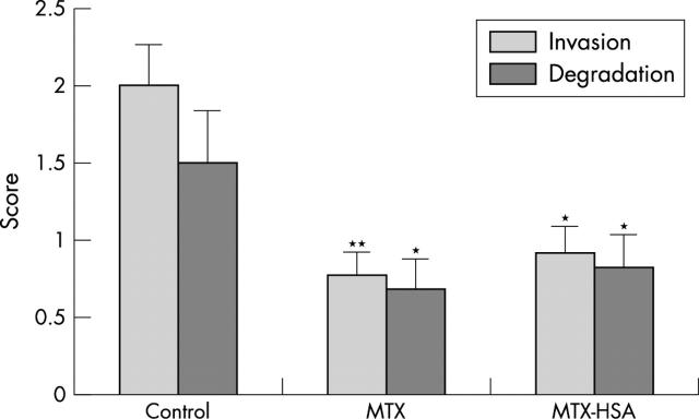
Results: The control group had a mean (SEM) score for cartilage invasion of RA SF of 2.0 (0.26) and for perichondrocytic cartilage degradation of 1.5 (0.34). In contrast, mice which received MTX showed a significantly reduced invasion (0.78 (0.14), p<0.01) and a reduction in perichondrocytic cartilage degradation scores (0.69 (0.2), p<0.05) in comparison with the control group. Mice treated with MTX-HSA also had significantly reduced scores for RA SF invasion into the cartilage (0.92 (0.41), p<0.05) and for cartilage degradation (0.83 (0.44), p<0.05) compared with controls. The effects of MTX and MTX-HSA were not significantly different between these two groups.
Conclusion: Treatment with MTX or MTX-HSA significantly ameliorates cartilage destruction in the SCID mouse model for human RA.
Full Text
The Full Text of this article is available as a PDF (88.8 KB).
Figure 1.
Treatment with MTX or albumin coupled MTX (MTX-HSA) suppresses cartilage invasion and perichondrocytic cartilage degradation in the SCID mice model of human RA. SCID mice were co-implanted subcutaneously with human cartilage and RA SF. Thereafter, they were treated with intraperitoneal injections twice weekly of 0.9% NaCl (control, n = 5), or 1 mg/kg MTX (n = 9), or 1 mg/kg MTX-HSA (n = 6). After 4 weeks of treatment, the implants were analysed histologically (fig 2), and cartilage invasion by SF as well as perichondrocytic cartilage degradation were scored (*p<0.05; **p<0.01; error bars represent SEM).
Figure 2.
H&E staining of co-transplanted RA SF and normal cartilage after an implantation time of 4 weeks from control mice (A; original magnification x800) and mice treated with either MTX (B; original magnification x1600) or MTX-HSA (original magnification x1600). In the control mice a deep invasion of the co-implanted RA SF into the cartilage can be seen (A), but in the mice which received treatment with either MTX or MTX-HSA the invasion is reduced (B) or even absent (C).


