Abstract
Objectives: To (a) develop further logically derived line drawing atlases (LDAs) for grading radiographic knee osteoarthritis (OA); and (b) determine which is superior using metrological criteria.
Methods: A series of LDAs (–3 to +3, –4 to +4, and –5 to +5) were produced by (a) incorporating additional grades for osteophyte and joint space width (JSW) above the 0–3 pilot LDA, over an equivalent range of disease; and (b) adding negative grades for JSW. 121 sets of bilateral knee radiographs (standing, anteroposterior plus flexed skyline), plus serial views of 68 tibiofemoral joints (TFJs) and 36 patellofemoral joints were scored twice by one observer for each LDA. Minimum JSW of 50 radiograph sets was directly measured and awarded a categorical grade dependent upon the boundaries of each LDA grade. Time taken to grade 30 randomly selected knee radiograph sets was measured.
Results: Intraobserver reproducibility was similar for all LDAs, (weighted κ: JSW = 0.85–0.87; osteophyte = 0.77–0.79), with no deterioration with increasing grades. Criterion validity favoured the –5 to +5 LDA, which was also quickest to use. All atlases showed similar responsiveness (standardised response mean: medial TFJ JSW = 0.78–0.83; medial femoral osteophyte = 0.61–0.73), with most sites compromised by small sample size, little change in score, and high variation between subjects.
Conclusions: A set of LDAs was created illustrating the full range of normality/abnormality likely to be encountered in a community study of knee pain or OA. Despite superior validity and equivalent reproducibility, improved responsiveness of the –5 to +5 LDA was not confirmed.
Full Text
The Full Text of this article is available as a PDF (367.7 KB).
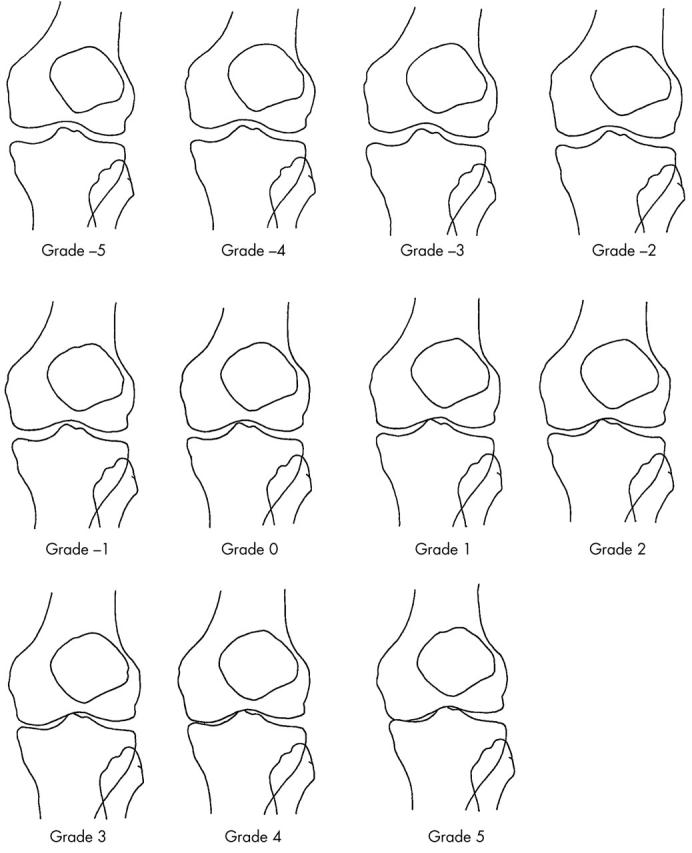
Figure 1.

–5 to +5 LDA: medial TFJ space width for women. Grades –5 to +5 (reduced size).
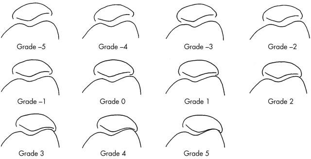
Figure 2.
–5 to +5 LDA: lateral PFJ space width for women. Grades –5 to +5 (reduced size).
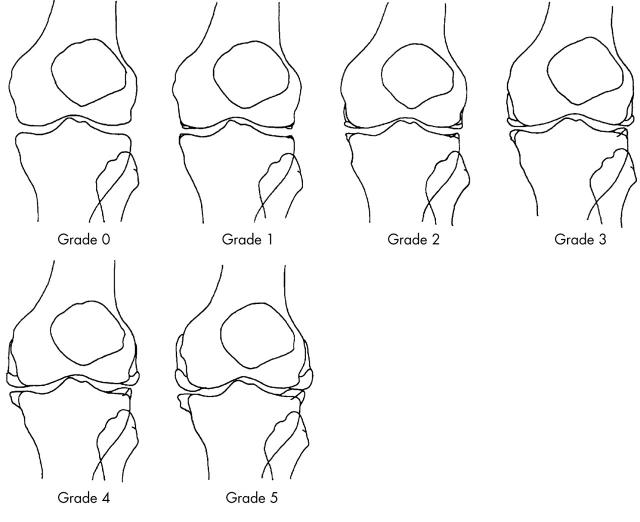
Figure 3.
–5 to +5 LDA: osteophyte in all tibiofemoral sites. Grades 0 to 5 (reduced size).
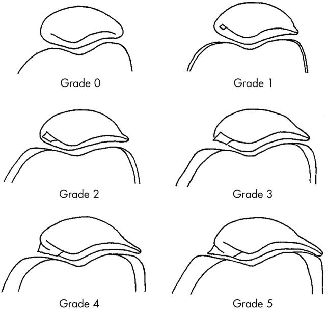
Figure 4.
–5 to +5 LDA: osteophyte in all patellofemoral sites. Grades 0 to 5 (reduced size).
Selected References
These references are in PubMed. This may not be the complete list of references from this article.
- Altman R. D., Fries J. F., Bloch D. A., Carstens J., Cooke T. D., Genant H., Gofton P., Groth H., McShane D. J., Murphy W. A. Radiographic assessment of progression in osteoarthritis. Arthritis Rheum. 1987 Nov;30(11):1214–1225. doi: 10.1002/art.1780301103. [DOI] [PubMed] [Google Scholar]
- Altman R. D., Hochberg M., Murphy W. A., Jr, Wolfe F., Lequesne M. Atlas of individual radiographic features in osteoarthritis. Osteoarthritis Cartilage. 1995 Sep;3 (Suppl A):3–70. [PubMed] [Google Scholar]
- Bland J. M., Altman D. G. Statistical methods for assessing agreement between two methods of clinical measurement. Lancet. 1986 Feb 8;1(8476):307–310. [PubMed] [Google Scholar]
- Braunstein E. M., Brandt K. D., Albrecht M. MRI demonstration of hypertrophic articular cartilage repair in osteoarthritis. Skeletal Radiol. 1990;19(5):335–339. doi: 10.1007/BF00193086. [DOI] [PubMed] [Google Scholar]
- Buckland-Wright J. C., Macfarlane D. G., Williams S. A., Ward R. J. Accuracy and precision of joint space width measurements in standard and macroradiographs of osteoarthritic knees. Ann Rheum Dis. 1995 Nov;54(11):872–880. doi: 10.1136/ard.54.11.872. [DOI] [PMC free article] [PubMed] [Google Scholar]
- Dacre J. E., Coppock J. S., Herbert K. E., Perrett D., Huskisson E. C. Development of a new radiographic scoring system using digital image analysis. Ann Rheum Dis. 1989 Mar;48(3):194–200. doi: 10.1136/ard.48.3.194. [DOI] [PMC free article] [PubMed] [Google Scholar]
- Hart D. J., Spector T. D. Kellgren & Lawrence grade 1 osteophytes in the knee--doubtful or definite? Osteoarthritis Cartilage. 2003 Feb;11(2):149–150. doi: 10.1053/joca.2002.0853. [DOI] [PubMed] [Google Scholar]
- Lanyon P., Jones A., Doherty M. Assessing progression of patellofemoral osteoarthritis: a comparison between two radiographic methods. Ann Rheum Dis. 1996 Dec;55(12):875–879. doi: 10.1136/ard.55.12.875. [DOI] [PMC free article] [PubMed] [Google Scholar]
- Lanyon P., O'Reilly S., Jones A., Doherty M. Radiographic assessment of symptomatic knee osteoarthritis in the community: definitions and normal joint space. Ann Rheum Dis. 1998 Oct;57(10):595–601. doi: 10.1136/ard.57.10.595. [DOI] [PMC free article] [PubMed] [Google Scholar]
- Laurin C. A., Dussault R., Levesque H. P. The tangential x-ray investigation of the patellofemoral joint: x-ray technique, diagnostic criteria and their interpretation. Clin Orthop Relat Res. 1979 Oct;(144):16–26. [PubMed] [Google Scholar]
- Leach R. E., Gregg T., Siber F. J. Weight-bearing radiography in osteoarthritis of the knee. Radiology. 1970 Nov;97(2):265–268. doi: 10.1148/97.2.265. [DOI] [PubMed] [Google Scholar]
- Ledingham J., Regan M., Jones A., Doherty M. Factors affecting radiographic progression of knee osteoarthritis. Ann Rheum Dis. 1995 Jan;54(1):53–58. doi: 10.1136/ard.54.1.53. [DOI] [PMC free article] [PubMed] [Google Scholar]
- Liang M. H., Fossel A. H., Larson M. G. Comparisons of five health status instruments for orthopedic evaluation. Med Care. 1990 Jul;28(7):632–642. doi: 10.1097/00005650-199007000-00008. [DOI] [PubMed] [Google Scholar]
- Maclure M., Willett W. C. Misinterpretation and misuse of the kappa statistic. Am J Epidemiol. 1987 Aug;126(2):161–169. doi: 10.1093/aje/126.2.161. [DOI] [PubMed] [Google Scholar]
- McAlindon T. E., Snow S., Cooper C., Dieppe P. A. Radiographic patterns of osteoarthritis of the knee joint in the community: the importance of the patellofemoral joint. Ann Rheum Dis. 1992 Jul;51(7):844–849. doi: 10.1136/ard.51.7.844. [DOI] [PMC free article] [PubMed] [Google Scholar]
- Nagaosa Y., Lanyon P., Doherty M. Characterisation of size and direction of osteophyte in knee osteoarthritis: a radiographic study. Ann Rheum Dis. 2002 Apr;61(4):319–324. doi: 10.1136/ard.61.4.319. [DOI] [PMC free article] [PubMed] [Google Scholar]
- Nagaosa Y., Mateus M., Hassan B., Lanyon P., Doherty M. Development of a logically devised line drawing atlas for grading of knee osteoarthritis. Ann Rheum Dis. 2000 Aug;59(8):587–595. doi: 10.1136/ard.59.8.587. [DOI] [PMC free article] [PubMed] [Google Scholar]
- Neame R. L., Carr A. J., Muir K., Doherty M. UK community prevalence of knee chondrocalcinosis: evidence that correlation with osteoarthritis is through a shared association with osteophyte. Ann Rheum Dis. 2003 Jun;62(6):513–518. doi: 10.1136/ard.62.6.513. [DOI] [PMC free article] [PubMed] [Google Scholar]
- Ravaud P., Ayral X., Dougados M. Radiologic progression of hip and knee osteoarthritis. Osteoarthritis Cartilage. 1999 Mar;7(2):222–229. doi: 10.1053/joca.1998.0155. [DOI] [PubMed] [Google Scholar]
- Ravaud P., Chastang C., Auleley G. R., Giraudeau B., Royant V., Amor B., Genant H. K., Dougados M. Assessment of joint space width in patients with osteoarthritis of the knee: a comparison of 4 measuring instruments. J Rheumatol. 1996 Oct;23(10):1749–1755. [PubMed] [Google Scholar]
- Ravaud P., Giraudeau B., Auleley G. R., Chastang C., Poiraudeau S., Ayral X., Dougados M. Radiographic assessment of knee osteoarthritis: reproducibility and sensitivity to change. J Rheumatol. 1996 Oct;23(10):1756–1764. [PubMed] [Google Scholar]
- Spector T. D., Hart D. J., Byrne J., Harris P. A., Dacre J. E., Doyle D. V. Definition of osteoarthritis of the knee for epidemiological studies. Ann Rheum Dis. 1993 Nov;52(11):790–794. doi: 10.1136/ard.52.11.790. [DOI] [PMC free article] [PubMed] [Google Scholar]
Associated Data
This section collects any data citations, data availability statements, or supplementary materials included in this article.


