Full Text
The Full Text of this article is available as a PDF (182.8 KB).
Figure 1.
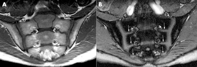
October 1996 (age 13 years and 4 months). (A) T1 weighted fast spin echo sequence depicts smoothly marginated sacroiliac joints of normal width. (B) Contrast enhanced T1 weighted gradient echo sequence without enhancement related to sacroiliitis. Of note, the growth plates of the sacral bone (arrows) are seen.
Figure 2.
February 1997 (age 13 years and 8 months). The boy presented with IBP that had persisted for 2 weeks. (A) T1 weighted fast spin echo sequence depicts a smoothly marginated right sacroiliac joint (star). The subchondral zone of the left iliac bone shows band-like hypointense formations with irregular borders (arrows), suggesting osteitis. (B) Corresponding contrast enhanced T1 weighted opposed phase gradient echo sequence showing pronounced enhancement within the joint space between the sacral and iliac cartilage (arrowheads) with continuous extension to the anterior and posterior joint capsule (arrows) and ligament attachments of the iliac bone. (C) Conventional radiograph shows normal joint contours (stars).
Figure 3.
February 1998 (age 14 years and 8 months). (A) T1 weighted fast spin echo image showing still normal width of the right sacroiliac joint. Band-like hypointensity in the iliac bone marrow, consistent with juxta-articular osteitis. The left anterior ilium shows a hypointense substrate (arrowhead), equivalent to an erosion and surrounding sclerosis. The posterior aspect shows an irregular hypointense configuration, indicating confluent erosions (double arrow) resulting in pseudodilatation of the joint. (B) Corresponding contrast enhanced T1 weighted opposed phase gradient echo sequence reveals pronounced contrast enhancement, both on the right in the area of the juxta-articular osteitis (arrowheads) and on the left in the area of the erosions (double arrows) and posterior joint capsule. (C) Conventional radiograph shows slight subchondral sclerosis of the left iliac bone (black arrow) with two suspected iliac erosions (arrowheads).
Figure 4.
March 2000 (age 16 years and 9 months). The patient presented without clinical symptoms at the sacroiliac joints. Nevertheless, follow up imaging demonstrates increasing chronic signs of bilateral sacroiliitis. (A) T1 weighted fast spin echo sequence visualises subchondral scleroses as irregular, partly linear, hypointensities (arrowheads). In addition, there is widening of the right and left joint space owing to beaded iliac erosions (arrows). (B) Corresponding contrast enhanced T1 weighted opposed phase gradient echo sequence showing minute enhancement in the joint space (arrowheads) and at the anterior capsule at the left side, indicative of only moderate inflammatory activity. (C) Anteroposterior radiograph depicting definite signs of bilateral sacroiliitis with sclerosis (black arrows) and widening of the joint space owing to confluent erosions.


