Full Text
The Full Text of this article is available as a PDF (774.5 KB).
Figure 1.
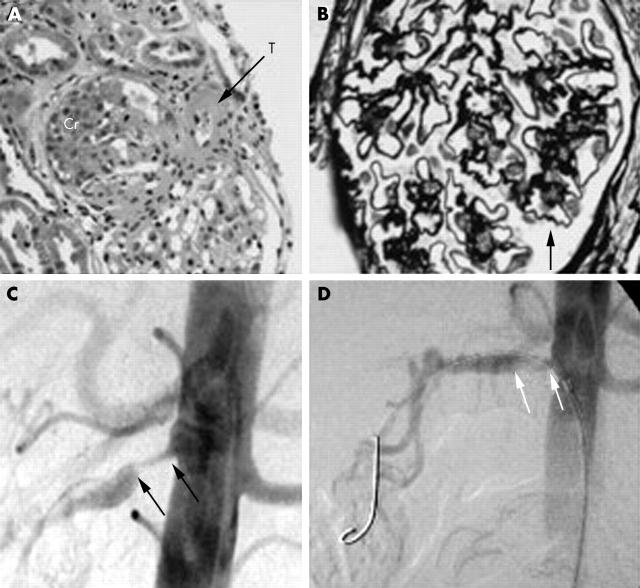
(A) Haematoxylin and eosin renal biopsy stain demonstrating a large cellular crescent (Cr) and intraluminal thrombus. (B) Periodic acid-Schiff/methanamine silver stain of renal biopsy specimen. The glomerular capillary walls and mesangial matrix are black. The folding/crenation (single arrow) suggests ischaemic contraction. (C) Renal angiogram demonstrating right renal artery ostial stenosis with post-stenotic dilatation (black arrows). Note the smooth non-atheromatous appearance of the aorta. (D) Percutaneous transluminal angioplasty of right renal artery stenosis (white arrows). A colour version of the figure can be seen at http://www.annrheumdis.com/supplemental.
Associated Data
This section collects any data citations, data availability statements, or supplementary materials included in this article.


