Abstract
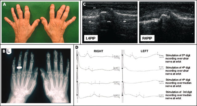
Case report: The patient developed arthritis mutilans in all digits of both hands with the exception of the left 4th finger, which had prior sensory denervation following traumatic nerve dissection. Plain radiography, ultrasonography and nerve conduction studies of the hands confirmed the absence of articular disease and sensory innervation in the left 4th digit.
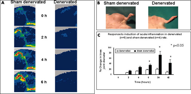
Methods: This relationship between joint innervation and joint inflammation was investigated experimentally by prior surgical sensory denervation of the medial aspect of the knee in six Wistar rats in which carrageenan induced arthritis was subsequently induced. Prior sensory denervation—with preservation of muscle function—prevented the development of inflammatory arthritis in the denervated knee.
Discussion: Observations in human and animal inflammatory arthritis suggest that regulatory neuroimmune pathways in the joint are an important mechanism that modulates the clinical expression of inflammatory arthritis.
Full Text
The Full Text of this article is available as a PDF (1.0 MB).
Figure 1.
(A) Swelling and deformity affecting most of the DIP and PIP joints of both hands. The left 4th digit is hypoplastic and has no evidence of joint inflammation or deformity. (B) Plain radiography demonstrates typical erosive joint damage and periostitis of PsA. The changes are most marked in the DIP joints and to a lesser extent in the PIP joints. (C) High resolution ultrasound of the left 4th PIP joint shows the clear bony outlines of the palmar aspects of the proximal phalanx (PP), PIP joint (PIPJ), and middle phalanx (MP) with the flexor tendon (FT) overlying. On the right, bony proliferation is present proximal (**) and distal to the PIP 4th joint. (D) Orthodromic nerve conduction studies from the right and left hands. From both hands stimulation of the 5th digit produced a normal ulnar sensory response. Stimulation of the right 4th digit produced a normal ulnar and median response. Stimulation of the left 4th digit produced no ulnar or median sensory response. In both hands stimulation of the 3rd digit produced a normal median sensory response. All waveforms were reproducible.
Figure 2.
(A) Perfusion images obtained from depilated rat knee joints scanned transcutaneously using a near-infrared (830 nm) laser Doppler imager. Acute inflammation was induced by injection of carrageenan/kaolin in both sham operated and denervated joints. Perfusion (flux) is colour coded with the lowest perfusion in dark blue, and the highest in red. Inflammatory hyperaemia fails to develop in the denervated knee. (B) At 24 hours after intra-articular carrageenan injection there is marked soft tissue swelling of the knee joint of the sham denervated animal but not in the denervated animal. (C) Changes in diameter of the knee joint over 48 hours demonstrate a rapid increase in swelling of the knee joint in the sham denervated animals, maximal at 24 hours, compared with the minimal change in joint diameter in the denervated animals. Student's t test with Bonferroni correction.


