Abstract
Objective: To identify classifiers in images obtained with sagittal laser optical tomography (SLOT) that can be used to distinguish between joints affected and not affected by synovitis.
Methods: 78 SLOT images of proximal interphalangeal joints II–IV from 13 patients with rheumatoid arthritis were compared with ultrasound (US) images and clinical examination (CE). SLOT images showing the spatial distribution of scattering and absorption coefficients within the joint cavity were generated. The means and standard errors for seven different classifiers (operator score and six quantitative measurements) were determined from SLOT images using CE and US as diagnostic references. For classifiers showing significant differences between affected and non-affected joints, sensitivities and specificities for various cut off parameters were obtained by receiver operating characteristic (ROC) analysis.
Results: For five classifiers used to characterise SLOT images the mean between affected and unaffected joints was statistically significant using US as diagnostic reference, but statistically significant for only one classifier with CE as reference. In general, high absorption and scattering coefficients in and around the joint cavity are indicative of synovitis. ROC analysis showed that the minimal absorption classifier yields the largest area under the curve (0.777; sensitivity and specificity 0.705 each) with US as diagnostic reference.
Conclusion: Classifiers in SLOT images have been identified that show statistically significant differences between joints with and without synovitis. It is possible to classify a joint as inflamed with SLOT, without the need for a reference measurement. Furthermore, SLOT based diagnosis of synovitis agrees better with US diagnosis than CE.
Full Text
The Full Text of this article is available as a PDF (496.5 KB).
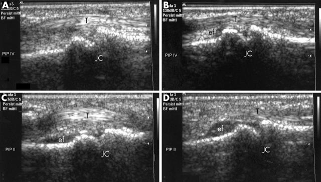
Figure 1.
Ultrasound images of PIP joint II (C, D) and IV (A, B) of patients with RA at different synovitis stages. T, tendon; JC, joint cavity. In all images, bone surface is without irregularities, no erosions are visible. Images are taken from the palmar side, and the left side of the image is nearer to, and the right side further from, the hand. Different extents of effusion (ef) can be seen in images B–D. Close to the synovial membrane, synovial proliferation can be detected in images C and D. The images were graded according to the degree of effusion and synovial hypertrophy using the adjusted semiquantitative score of Szkudlarek et al15: (A) grade 0 = none; (B) grade 1 = minimal; (C) grade 2 = moderate; and (D) grade 3 = extensive; the degree of inflammation was interpreted by effusion and synovitis.
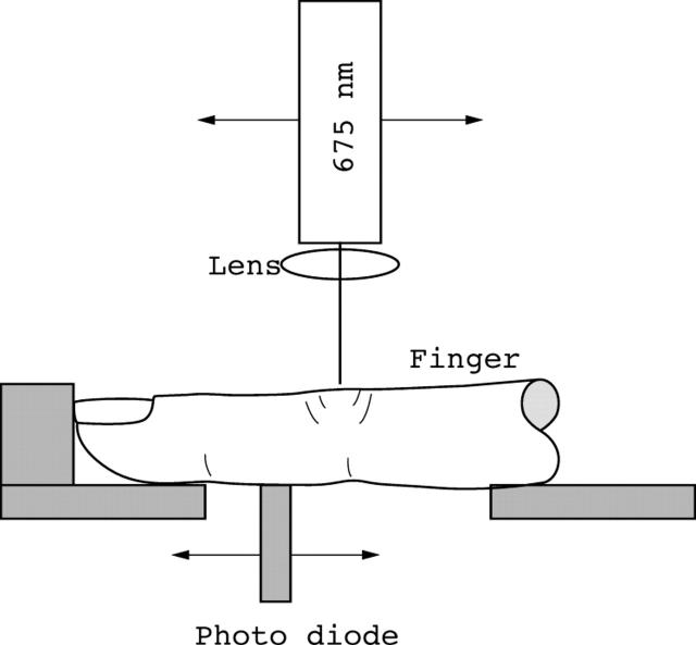
Figure 2.
Experimental setup for sagittal joint imaging. The laser is positioned above and a photodetector is placed below the finger joint to be examined. Both, detector and diode laser are attached to stepping motor driven translation stages that permit independent control of the position of the laser diode and photodetector relative to the joint. Detector and laser are connected to a personal computer, where data are collected.
Figure 3.
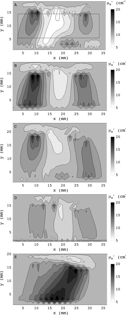
Reconstructed cross sections of the scattering coefficient for three different fingers typical for (A) category 1—definitely no synovitis; (B) category 2—probably no synovitis; (C) category 3—possibly synovitis; (D) category 4—probably synovitis; and (E) category 5—definitely synovitis. The fingertip is located to the right of the images that show a 36 mm wide section of the finger with the joint cavity located approximately in the centre. The rectangle in fig 3A indicates the region for which Min(µs), Min(µa), Max(µs), Max(µa), Min(µs)/Max(µs), and Min(µa)/Max(µa), were calculated.
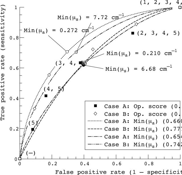
Figure 4.
ROC curves with ultrasound scores of 0—unaffected and 3—affected as diagnostic reference (case B) compared with ROC curves with ultrasound scores of (0, 1)—affected and (2,3)—unaffected as diagnostic reference (case A). The numbers in brackets are the area under the curve (AUC). Also given for each curve are the cut off values that result in the largest Youden index.
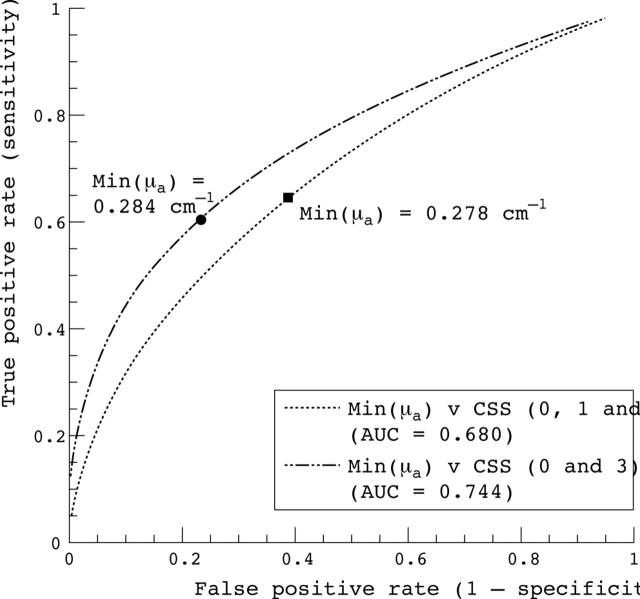
Figure 5.
ROC curves for Min(µa) classifier with clinical scores as diagnostic reference. The two cut off values identify points on the curve for which the Youden index is maximal.
Selected References
These references are in PubMed. This may not be the complete list of references from this article.
- Arnett F. C., Edworthy S. M., Bloch D. A., McShane D. J., Fries J. F., Cooper N. S., Healey L. A., Kaplan S. R., Liang M. H., Luthra H. S. The American Rheumatism Association 1987 revised criteria for the classification of rheumatoid arthritis. Arthritis Rheum. 1988 Mar;31(3):315–324. doi: 10.1002/art.1780310302. [DOI] [PubMed] [Google Scholar]
- Backhaus M., Kamradt T., Sandrock D., Loreck D., Fritz J., Wolf K. J., Raber H., Hamm B., Burmester G. R., Bollow M. Arthritis of the finger joints: a comprehensive approach comparing conventional radiography, scintigraphy, ultrasound, and contrast-enhanced magnetic resonance imaging. Arthritis Rheum. 1999 Jun;42(6):1232–1245. doi: 10.1002/1529-0131(199906)42:6<1232::AID-ANR21>3.0.CO;2-3. [DOI] [PubMed] [Google Scholar]
- Backhaus M., Schmidt W. A., Mellerowicz H., Bohl-Bühler M., Banzer D., Braun J., Sattler H., Hauer R-W, Arbeitskreis "bildgebende Diagnostik in der Rheumatologie" des Regionalen Rheumazentrums Berlin, e.V Technik und Stellenwert der Arthrosonographie in der rheumatologischen Diagnostik -- Teil 6: Sonographie der Hand- und Fingergelenke. Z Rheumatol. 2002 Dec;61(6):674–687. doi: 10.1007/s00393-002-0386-6. [DOI] [PubMed] [Google Scholar]
- Beuthan J., Cappius H. J., Hielscher A., Hopf M., Klose A., Netz U. Erste Untersuchungen zur Anwendung der linearen Signalübertragungstheorie in der Auswertung diaphanoskopischer Untersuchungen am Beispiel der Rheumadiagnostik. Biomed Tech (Berl) 2001 Nov;46(11):298–303. doi: 10.1515/bmte.2001.46.11.298. [DOI] [PubMed] [Google Scholar]
- Hermann Kay-Geert A., Backhaus Marina, Schneider Udo, Labs Karsten, Loreck Dieter, Zühlsdorf Svenda, Schink Tania, Fischer Thomas, Hamm Bernd, Bollow Matthias. Rheumatoid arthritis of the shoulder joint: comparison of conventional radiography, ultrasound, and dynamic contrast-enhanced magnetic resonance imaging. Arthritis Rheum. 2003 Dec;48(12):3338–3349. doi: 10.1002/art.11349. [DOI] [PubMed] [Google Scholar]
- Hielscher A. H., Bluestone A. Y., Abdoulaev G. S., Klose A. D., Lasker J., Stewart M., Netz U., Beuthan J. Near-infrared diffuse optical tomography. Dis Markers. 2002;18(5-6):313–337. doi: 10.1155/2002/164252. [DOI] [PMC free article] [PubMed] [Google Scholar]
- Hielscher A. H., Klose A. D., Hanson K. M. Gradient-based iterative image reconstruction scheme for time-resolved optical tomography. IEEE Trans Med Imaging. 1999 Mar;18(3):262–271. doi: 10.1109/42.764902. [DOI] [PubMed] [Google Scholar]
- Hielscher Andreas H., Klose Alexander D., Scheel Alexander K., Moa-Anderson Bryte, Backhaus Marina, Netz Uwe, Beuthan Jürgen. Sagittal laser optical tomography for imaging of rheumatoid finger joints. Phys Med Biol. 2004 Apr 7;49(7):1147–1163. doi: 10.1088/0031-9155/49/7/005. [DOI] [PubMed] [Google Scholar]
- Kane David, Balint Peter V., Sturrock Roger D. Ultrasonography is superior to clinical examination in the detection and localization of knee joint effusion in rheumatoid arthritis. J Rheumatol. 2003 May;30(5):966–971. [PubMed] [Google Scholar]
- Kim J. M., Weisman M. H. When does rheumatoid arthritis begin and why do we need to know? Arthritis Rheum. 2000 Mar;43(3):473–484. doi: 10.1002/1529-0131(200003)43:3<473::AID-ANR1>3.0.CO;2-A. [DOI] [PubMed] [Google Scholar]
- Klose A. D., Hielscher A. H. Iterative reconstruction scheme for optical tomography based on the equation of radiative transfer. Med Phys. 1999 Aug;26(8):1698–1707. doi: 10.1118/1.598661. [DOI] [PubMed] [Google Scholar]
- Lassere Marissa, McQueen Fiona, Østergaard Mikkel, Conaghan Philip, Shnier Ron, Peterfy Charles, Klarlund Mette, Bird Paul, O'Connor Philip, Stewart Neal. OMERACT Rheumatoid Arthritis Magnetic Resonance Imaging Studies. Exercise 3: an international multicenter reliability study using the RA-MRI Score. J Rheumatol. 2003 Jun;30(6):1366–1375. [PubMed] [Google Scholar]
- Metz CE, Pan X. "Proper" Binormal ROC Curves: Theory and Maximum-Likelihood Estimation. J Math Psychol. 1999 Mar;43(1):1–33. doi: 10.1006/jmps.1998.1218. [DOI] [PubMed] [Google Scholar]
- Peterfy Charles G. New developments in imaging in rheumatoid arthritis. Curr Opin Rheumatol. 2003 May;15(3):288–295. doi: 10.1097/00002281-200305000-00017. [DOI] [PubMed] [Google Scholar]
- Prapavat V., Runge W., Mans J., Krause A., Beuthan J., Müller G. Entwicklung eines Fingergelenkphantoms zur optischen Simulation früher entzündlich-rheumatischer Veränderungen. Biomed Tech (Berl) 1997 Nov;42(11):319–326. doi: 10.1515/bmte.1997.42.11.319. [DOI] [PubMed] [Google Scholar]
- Qvistgaard E., Røgind H., Torp-Pedersen S., Terslev L., Danneskiold-Samsøe B., Bliddal H. Quantitative ultrasonography in rheumatoid arthritis: evaluation of inflammation by Doppler technique. Ann Rheum Dis. 2001 Jul;60(7):690–693. doi: 10.1136/ard.60.7.690. [DOI] [PMC free article] [PubMed] [Google Scholar]
- Scheel Alexander K., Krause Andreas, Rheinbaben Ingolf Mesecke-Von, Metzger Georg, Rost Helmut, Tresp Volker, Mayer Peter, Reuss-Borst Monika, Müller Gerhard A. Assessment of proximal finger joint inflammation in patients with rheumatoid arthritis, using a novel laser-based imaging technique. Arthritis Rheum. 2002 May;46(5):1177–1184. doi: 10.1002/art.10226. [DOI] [PubMed] [Google Scholar]
- Schmidt W. A., Völker L., Zacher J., Schläfke M., Ruhnke M., Gromnica-Ihle E. Colour Doppler ultrasonography to detect pannus in knee joint synovitis. Clin Exp Rheumatol. 2000 Jul-Aug;18(4):439–444. [PubMed] [Google Scholar]
- Schwaighofer Anton, Tresp Volker, Mayer Peter, Krause Andreas, Beuthan Jürgen, Rost Helmut, Metzger Georg, Müller Gerhard A., Scheel Alexander K. Classification of rheumatoid joint inflammation based on laser imaging. IEEE Trans Biomed Eng. 2003 Mar;50(3):375–382. doi: 10.1109/TBME.2003.808827. [DOI] [PubMed] [Google Scholar]
- Scott D. L., Coulton B. L., Popert A. J. Long term progression of joint damage in rheumatoid arthritis. Ann Rheum Dis. 1986 May;45(5):373–378. doi: 10.1136/ard.45.5.373. [DOI] [PMC free article] [PubMed] [Google Scholar]
- Szkudlarek M., Court-Payen M., Strandberg C., Klarlund M., Klausen T., Ostergaard M. Power Doppler ultrasonography for assessment of synovitis in the metacarpophalangeal joints of patients with rheumatoid arthritis: a comparison with dynamic magnetic resonance imaging. Arthritis Rheum. 2001 Sep;44(9):2018–2023. doi: 10.1002/1529-0131(200109)44:9<2018::AID-ART350>3.0.CO;2-C. [DOI] [PubMed] [Google Scholar]
- Taylor Peter C. Anti-TNFalpha therapy for rheumatoid arthritis: an update. Intern Med. 2003 Jan;42(1):15–20. [PubMed] [Google Scholar]
- Terslev L., Torp-Pedersen S., Savnik A., von der Recke P., Qvistgaard E., Danneskiold-Samsøe B., Bliddal H. Doppler ultrasound and magnetic resonance imaging of synovial inflammation of the hand in rheumatoid arthritis: a comparative study. Arthritis Rheum. 2003 Sep;48(9):2434–2441. doi: 10.1002/art.11245. [DOI] [PubMed] [Google Scholar]
- Verstappen Suzan M. M., Jacobs Johannes W. G., Bijlsma Johannes W. J., Heurkens Anton H. M., van Booma-Frankfort Christina, Borg Evert Jan ter, Hofman Dick M., van der Veen Maaike J., Utrecht Arthritis Cohort Study Group Five-year followup of rheumatoid arthritis patients after early treatment with disease-modifying antirheumatic drugs versus treatment according to the pyramid approach in the first year. Arthritis Rheum. 2003 Jul;48(7):1797–1807. doi: 10.1002/art.11170. [DOI] [PubMed] [Google Scholar]
- Wakefield R. J., Kong K. O., Conaghan P. G., Brown A. K., O'Connor P. J., Emery P. The role of ultrasonography and magnetic resonance imaging in early rheumatoid arthritis. Clin Exp Rheumatol. 2003 Sep-Oct;21(5 Suppl 31):S42–S49. [PubMed] [Google Scholar]
- Xu Yong, Iftimia Nicusor, Jiang Huabei, Key L. Lyndon, Bolster Marcy B. Three-dimensional diffuse optical tomography of bones and joints. J Biomed Opt. 2002 Jan;7(1):88–92. doi: 10.1117/1.1427336. [DOI] [PubMed] [Google Scholar]
- YOUDEN W. J. Index for rating diagnostic tests. Cancer. 1950 Jan;3(1):32–35. doi: 10.1002/1097-0142(1950)3:1<32::aid-cncr2820030106>3.0.co;2-3. [DOI] [PubMed] [Google Scholar]


