Abstract
Case report: A patient presented with severe treatment resistant PVNS of the right knee joint. Several conventional treatment regimens, including open surgical synovectomy and intra-articular injections of yttrium-90 (90Y) failed to control the disease. After finding marked tumour necrosis factor α (TNFα) expression in arthroscopic synovial tissue samples, treatment with an anti-TNFα monoclonal antibody (infliximab) at a dose of 5 mg/kg was started. Additional courses with the same dose given 2, 6, 14, and 20 weeks later, and bimonthly thereafter up to 54 weeks, controlled the signs and symptoms. Immunohistological analysis at follow up identified a marked reduction in macrophage numbers and TNFα expression in the synovium.
Discussion: This is probably the first case which describes treatment with TNFα blockade of PVNS in a patient who is refractory to conventional treatment. It provides the rationale for larger controlled studies to elucidate further the efficacy of TNFα blockade treatment in refractory PVNS.
Full Text
The Full Text of this article is available as a PDF (147.1 KB).
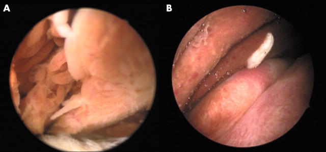
Figure 1.
Macroscopic examination of the synovium before (A) and after (B) 20 weeks of treatment with anti-TNFα. (A) Hypertrophied synovium with villous transformation and haemosiderin deposition. (B) Reduction of hypertrophied synovium and haemosiderin deposition.
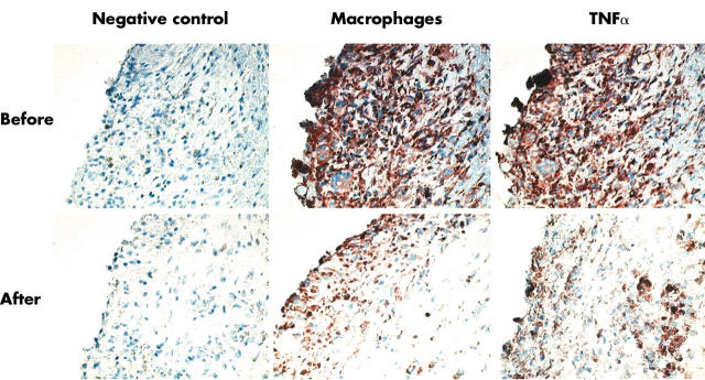
Figure 2.
Pathological specimen of pigmented villonodular synovitis before and after 20 weeks of treatment with anti-TNFα. Before treatment: classic histological features were seen, with pigment depositions and an impressive infiltrate, mainly consisting of macrophages with an abundant presence of TNFα, and immunohistochemical staining for macrophages and TNFα at baseline. After treatment: a dramatic reduction in cellularity, number of macrophages, estimated pigment, and TNFα after 20 weeks of treatment with anti-TNFα was seen.


