Full Text
The Full Text of this article is available as a PDF (200.1 KB).
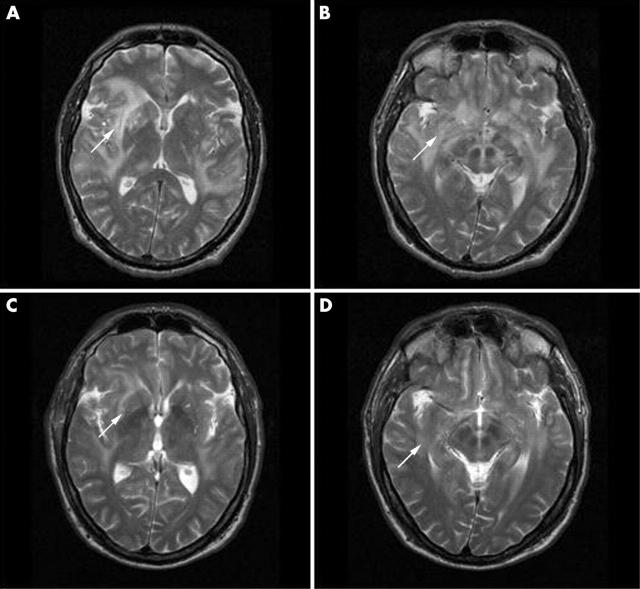
Figure 1.
(A, B) Axial T2 weighted fast spin echo MR images of the brain show increased signal intensity in the bilateral white matter and basal ganglia (arrows). (C, D) Follow up MRI shows a dramatic improvement of the white matter and basal ganglia abnormalities only 8 weeks after starting treatment.
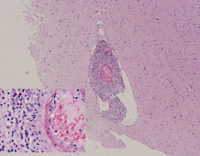
Figure 2.
A cerebral biopsy specimen was taken from the right parietal hemisphere, leptomeninx, and cortex (original magnification x200 and x40). Severe histopathological signs of cerebral vasculitis in the cortex and leptomeninx can be seen, consisting of a diffuse mononuclear cell infiltrate through the whole vessel wall with fibrinoid necrosis in the presence of a normal cerebral angiogram of the same hemisphere.


