Abstract
Methods: The composition of 10 tendon samples from patients with a prior history of painful Achilles tendinosis and 10 samples from patients with spontaneously ruptured tendons but no previous pain was compared by immunohistochemistry and conventional histology.
Results: The presence of granulation tissue was shown in 8/10 cases of Achilles tendinosis. Nociceptive substance P (SP) positive nerve fibres were significantly increased, and an inflammatory infiltration comprising B and T lymphocytes was found. Additionally, small foci with iron positive haemosiderophages, indicating prior microtraumatic events, were found in 6/10 samples. None of the spontaneously ruptured tendons contained granulation tissue or haemosiderophages. Inflammatory infiltration in these patients consisted almost exclusively of granulocytes and SP positive nerve fibres were decreased. The density of sympathetic nerve fibres did not differ in the two conditions.
Conclusion: Achilles tendinosis is associated with the presence of granulation tissue, haemosiderophages, and SP positive nerve fibres, which may transmit the clinically pertinent pain. Achilles tendinosis may be caused by repeated microtraumata with ensuing organisation that is accompanied by sprouting of nociceptive SP positive nerve fibres.
Full Text
The Full Text of this article is available as a PDF (98.5 KB).
Figure 1.
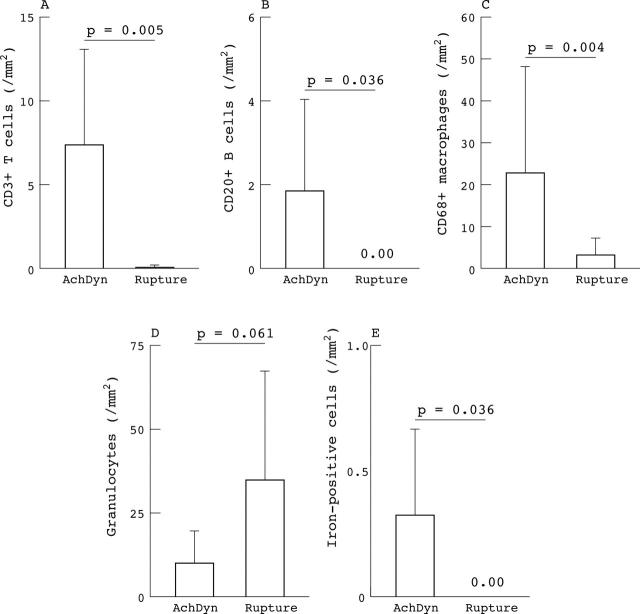
Determination of cell types in painful Achilles tendinosis and ruptured Achilles tendon. The numbers of (A) CD3+ and (B) CD20+ lymphocytes and (C) CD68+ macrophages were determined by immunohistochemistry in 10 tendons from patients with Achilles tendinosis and in 10 tendons from patients with spontaneously ruptured tendons. The number of (D) granulocytes was determined on haematoxylin and eosin stained sections and the number of (E) iron positive haemosiderophages was determined in sections stained by Turnbull's acid ferrocyanide reaction. The number of all cells was averaged from 10 high power fields and expressed as the number of cells/mm2. AchDyn, painful Achilles tendinosis.
Figure 3.
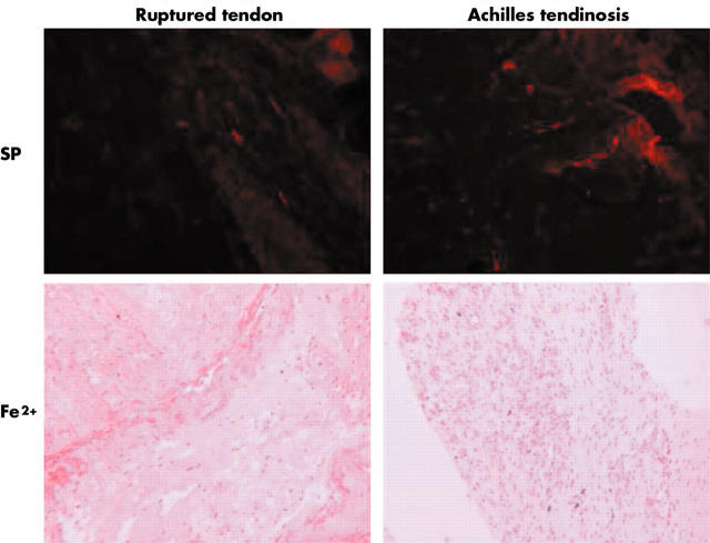
Histological sections of specimens from patients with ruptured tendons and from patients with tendinosis stained for substance P by immunofluorescence (SP, top row, original magnification x400) and stained by Turnbull's acid ferrocyanide reaction to detect haemosiderophages (Fe2+, bottom row, original magnification x5). Note that there are more SP positive nerve fibres visible in the tendinosis specimen than in the ruptured tendon. Haemosiderophages which are discernible as dark blue spots and small blood vessels were only found in the tendinosis specimen, not in the ruptured tendon.
Figure 2.
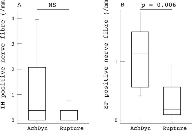
Comparison of sympathetic and nociceptive nerve fibres in painful tendinosis and ruptured tendon. The numbers of TH positive nerve fibres (A) and SP positive nerve fibres (B) were determined by immunofluorescent histochemistry in 10 tendons from patients with Achilles tendinosis (AchDyn) and 10 tendons from patients with spontaneously ruptured tendons. The number of nerve fibres was averaged from 17 high power fields and expressed as the number of fibres/mm2. Box plots give the 10th, 25th, 75th, and 90th centiles. The median is given as a horizontal line within the box. SP, substance P; TH, tyrosine hydroxylase (sympathetic nerve fibres).


