Full Text
The Full Text of this article is available as a PDF (101.6 KB).
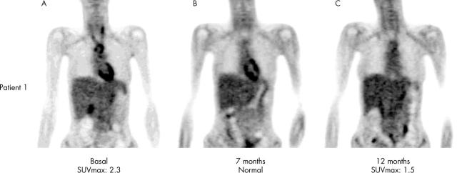
Figure 1.
(A) Hypermetabolism is detected in the brachiocephalic trunk, left carotid artery, and thoracic aorta. (B) Normal thoracic metabolism (unspecific bowel uptake can be seen in the abdominal area). (C) Hypermetabolism is detected in the aortic arch and the proximal third of both carotid arteries.
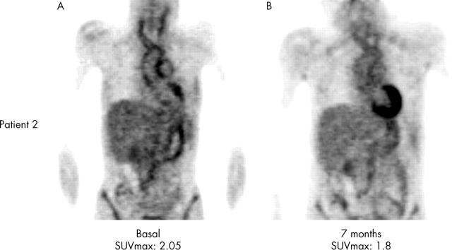
Figure 2.
Figures 2 (A and B) PET shows metabolism in the thoracic aorta, carotids, brachiocephalic trunk and pulmonary trunk.


