Abstract
BACKGROUND/AIMS/METHODS—During hepatic vein catheterisation, in addition to measurement of hepatic venous pressure gradient (HVPG), iodine wedged retrograde portography can be easily obtained. However, it rarely allows correct visualisation of the portal vein. Recently, CO2 has been suggested to allow better angiographic demonstration of the portal vein than iodine. In this study we investigated the efficacy of CO2 compared with iodinated contrast medium for portal vein imaging and its role in the evaluation of portal hypertension in a series of 100 patients undergoing hepatic vein catheterisation, 71 of whom had liver cirrhosis. RESULTS—In the overall series, CO2 venography was markedly superior to iodine, allowing correct visualisation of the different segments of the portal venous system. In addition, CO2, but not iodine, visualised portal-systemic collaterals in 34 patients. In cirrhosis, non-visualisation of the portal vein on CO2 venography occurred in 11 cases; four had portal vein thrombosis and five had communications between different hepatic veins. Among non-cirrhotics, lack of portal vein visualisation had a 90% sensitivity, 88% specificity, 94% negative predictive value, and 83% positive predictive value in the diagnosis of pre-sinusoidal portal hypertension. CONCLUSIONS—Visualisation of the venous portal system by CO2 venography is markedly superior to iodine. The use of CO2 wedged portography is a useful and safe complementary procedure during hepatic vein catheterisation which may help to detect portal thrombosis. Also, lack of demonstration of the portal vein in non-cirrhotic patients strongly suggests the presence of pre-sinusoidal portal hypertension. Keywords: portal thrombosis; liver cirrhosis; idiopathic portal hypertension; splanchnic haemodynamics
Full Text
The Full Text of this article is available as a PDF (173.8 KB).
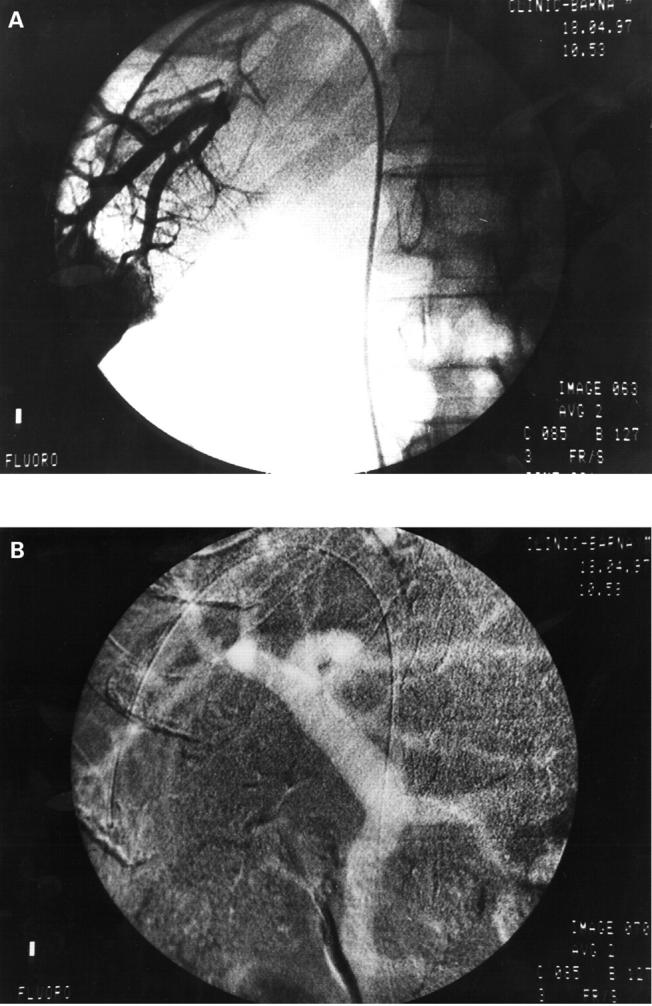
Figure 1 .

Iodine venography (A) allowed visualisation of the distal branches of the intrahepatic portal vein while CO2 venography (B) allowed opacification of the whole spleno-mesenteric-portal system.
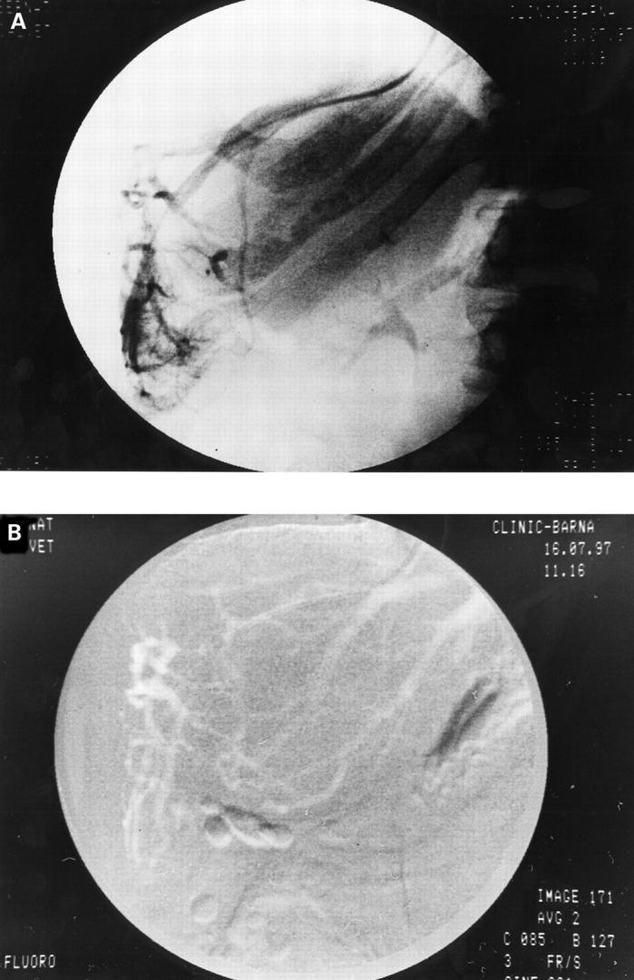
Figure 2 .
CO2 venography allowed visualisation of the whole spleno-mesenteric-portal system, including the extrahepatic collaterals.
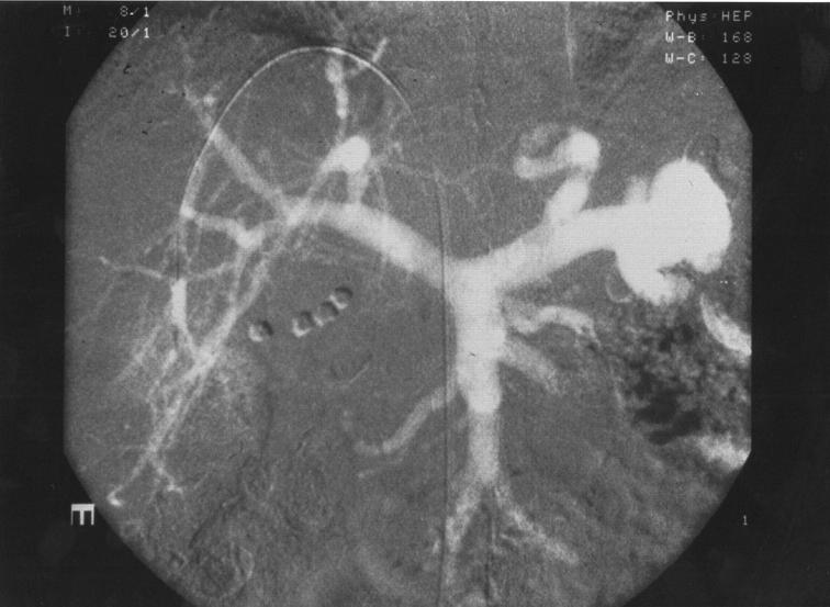
Figure 3 .

Idiopathic portal hypertension. The portal vein is not opacified either on iodine (A) or CO2 (B) venography. Several veno-venous communications are seen.
Selected References
These references are in PubMed. This may not be the complete list of references from this article.
- Boyer J. L., Sen Gupta K. P., Biswas S. K., Pal N. C., Basu Mallick K. C., Iber F. L., Basu A. K. Idiopathic portal hypertension. Comparison with the portal hypertension of cirrhosis and extrahepatic portal vein obstruction. Ann Intern Med. 1967 Jan;66(1):41–68. doi: 10.7326/0003-4819-66-1-41. [DOI] [PubMed] [Google Scholar]
- Dockray K. T., Burns R. R., Moore J. Capnohepatography: intravenous retrograde gas angiography of liver veins with carbon dioxide. Radiology. 1965 Oct;85(4):740–742. doi: 10.1148/85.4.740. [DOI] [PubMed] [Google Scholar]
- Feu F., García-Pagán J. C., Bosch J., Luca A., Terés J., Escorsell A., Rodés J. Relation between portal pressure response to pharmacotherapy and risk of recurrent variceal haemorrhage in patients with cirrhosis. Lancet. 1995 Oct 21;346(8982):1056–1059. doi: 10.1016/s0140-6736(95)91740-3. [DOI] [PubMed] [Google Scholar]
- Futagawa S., Fukazawa M., Musha H., Isomatsu T., Koyama K., Ito T., Horisawa M., Nakayama S., Sugiura M., Kameda H. Hepatic venography in noncirrhotic idiopathic portal hypertension. Comparison with cirrhosis of the liver. Radiology. 1981 Nov;141(2):303–309. doi: 10.1148/radiology.141.2.7291551. [DOI] [PubMed] [Google Scholar]
- Garcia-Pagán J. C., Navasa M., Bosch J., Bru C., Pizcueta P., Rodés J. Enhancement of portal pressure reduction by the association of isosorbide-5-mononitrate to propranolol administration in patients with cirrhosis. Hepatology. 1990 Feb;11(2):230–238. doi: 10.1002/hep.1840110212. [DOI] [PubMed] [Google Scholar]
- Garcia-Tsao G., Groszmann R. J., Fisher R. L., Conn H. O., Atterbury C. E., Glickman M. Portal pressure, presence of gastroesophageal varices and variceal bleeding. Hepatology. 1985 May-Jun;5(3):419–424. doi: 10.1002/hep.1840050313. [DOI] [PubMed] [Google Scholar]
- Groszmann R. J., Atterbury C. E. The pathophysiology of portal hypertension: a basis for classification. Semin Liver Dis. 1982 Aug;2(3):177–186. doi: 10.1055/s-2008-1040707. [DOI] [PubMed] [Google Scholar]
- Groszmann R. J., Bosch J., Grace N. D., Conn H. O., Garcia-Tsao G., Navasa M., Alberts J., Rodes J., Fischer R., Bermann M. Hemodynamic events in a prospective randomized trial of propranolol versus placebo in the prevention of a first variceal hemorrhage. Gastroenterology. 1990 Nov;99(5):1401–1407. doi: 10.1016/0016-5085(90)91168-6. [DOI] [PubMed] [Google Scholar]
- Groszmann R. J. The hepatic venous pressure gradient: has the time arrived for its application in clinical practice? Hepatology. 1996 Sep;24(3):739–741. doi: 10.1002/hep.510240345. [DOI] [PubMed] [Google Scholar]
- Hawkins I. F., Jr, Wilcox C. S., Kerns S. R., Sabatelli F. W. CO2 digital angiography: a safer contrast agent for renal vascular imaging? Am J Kidney Dis. 1994 Oct;24(4):685–694. doi: 10.1016/s0272-6386(12)80232-0. [DOI] [PubMed] [Google Scholar]
- Kerns S. R., Hawkins I. F., Jr Carbon dioxide digital subtraction angiography: expanding applications and technical evolution. AJR Am J Roentgenol. 1995 Mar;164(3):735–741. doi: 10.2214/ajr.164.3.7863904. [DOI] [PubMed] [Google Scholar]
- Krasny R., Hollmann J. P., Günther R. W. Erste Erfahrungen mit CO2 als gasförmiges Kontrastmittel in der DSA. Rofo. 1987 Apr;146(4):450–454. doi: 10.1055/s-2008-1048519. [DOI] [PubMed] [Google Scholar]
- Lebrec D., Degott C., Rueff B., Benhamou J. P. Transvenous (transjugular) liver biopsy. An experience based on 100 biopsies. Am J Dig Dis. 1978 Apr;23(4):302–304. doi: 10.1007/BF01072410. [DOI] [PubMed] [Google Scholar]
- Merkel C., Bolognesi M., Bellon S., Zuin R., Noventa F., Finucci G., Sacerdoti D., Angeli P., Gatta A. Prognostic usefulness of hepatic vein catheterization in patients with cirrhosis and esophageal varices. Gastroenterology. 1992 Mar;102(3):973–979. doi: 10.1016/0016-5085(92)90185-2. [DOI] [PubMed] [Google Scholar]
- Novak D., Bützow G. H., Becker K. Hepatic occlusion venography with a balloon catheter in portal hypertension. Radiology. 1977 Mar;122(3):623–628. doi: 10.1148/122.3.623. [DOI] [PubMed] [Google Scholar]
- Okuda K., Nakashima T., Okudaira M., Kage M., Aida Y., Omata M., Musha H., Futagawa S., Sugiura M., Kameda H. Anatomical basis of hepatic venographic alterations in idiopathic portal hypertension. Liver. 1981 Dec;1(4):255–263. doi: 10.1111/j.1600-0676.1981.tb00041.x. [DOI] [PubMed] [Google Scholar]
- Okuda K., Ohnishi K., Kimura K., Matsutani S., Sumida M., Goto N., Musha H., Takashi M., Suzuki N., Shinagawa T. Incidence of portal vein thrombosis in liver cirrhosis. An angiographic study in 708 patients. Gastroenterology. 1985 Aug;89(2):279–286. doi: 10.1016/0016-5085(85)90327-0. [DOI] [PubMed] [Google Scholar]
- Okuda K., Takayasu K., Matsutani S. Angiography in portal hypertension. Gastroenterol Clin North Am. 1992 Mar;21(1):61–83. [PubMed] [Google Scholar]
- RAPPAPORT A. M., HOLMES R. B., STOLBERG H. O., MCINTYRE J. L., BAIRD R. J. HEPATIC VENOGRAPHY. Gastroenterology. 1964 Feb;46:115–127. [PubMed] [Google Scholar]
- SCHLANT R. C., GALAMBOS J. T., SHUFORD W. H., RAWLS W. J., WINTER T. S., 3rd, EDWARDS F. K. THE CLINICAL USEFULNESS OF WEDGE HEPATIC VENOGRAPHY. Am J Med. 1963 Sep;35:343–349. doi: 10.1016/0002-9343(63)90176-1. [DOI] [PubMed] [Google Scholar]
- Sullivan K. L., Bonn J., Shapiro M. J., Gardiner G. A. Venography with carbon dioxide as a contrast agent. Cardiovasc Intervent Radiol. 1995 May-Jun;18(3):141–145. doi: 10.1007/BF00204138. [DOI] [PubMed] [Google Scholar]
- The value of Doppler US in the study of hepatic hemodynamics. Consensus conference (Bologna, Italy, 12 September, 1989). J Hepatol. 1990 May;10(3):353–355. doi: 10.1016/0168-8278(90)90146-i. [DOI] [PubMed] [Google Scholar]
- Trejo R., Alvarez W., García-Pagán J. C., Feu F., Escorsell A., Bruguera M., Bosch J., Rodés J. Aplicabilidad y rentabilidad diagnóstica de la biopsia hepática transyugular. Med Clin (Barc) 1996 Oct 26;107(14):521–523. [PubMed] [Google Scholar]
- Triger D. R. Extra hepatic portal venous obstruction. Gut. 1987 Oct;28(10):1193–1197. doi: 10.1136/gut.28.10.1193. [DOI] [PMC free article] [PubMed] [Google Scholar]
- Weaver F. A., Pentecost M. J., Yellin A. E., Davis S., Finck E., Teitelbaum G. Clinical applications of carbon dioxide/digital subtraction arteriography. J Vasc Surg. 1991 Feb;13(2):266–273. [PubMed] [Google Scholar]
- Wilhelm K., Textor J., Strunk H., Brensing K. A., Schüller H., Schild H. Kohlendioxid (CO2) als Kontrastmittel zur Neuanlage und Kontrolle von TIPS. Rofo. 1997 Mar;166(3):238–242. doi: 10.1055/s-2007-1015416. [DOI] [PubMed] [Google Scholar]
- Williams D. M., Cho K. J., Aisen A. M., Eckhauser F. E. Portal hypertension evaluated by MR imaging. Radiology. 1985 Dec;157(3):703–706. doi: 10.1148/radiology.157.3.4059557. [DOI] [PubMed] [Google Scholar]
- Yusuf S. W., Whitaker S. C., Hinwood D., Henderson M. J., Gregson R. H., Wenham P. W., Hopkinson B. R., Makin G. S. Carbon dioxide: an alternative to iodinated contrast media. Eur J Vasc Endovasc Surg. 1995 Aug;10(2):156–161. doi: 10.1016/s1078-5884(05)80106-6. [DOI] [PubMed] [Google Scholar]


