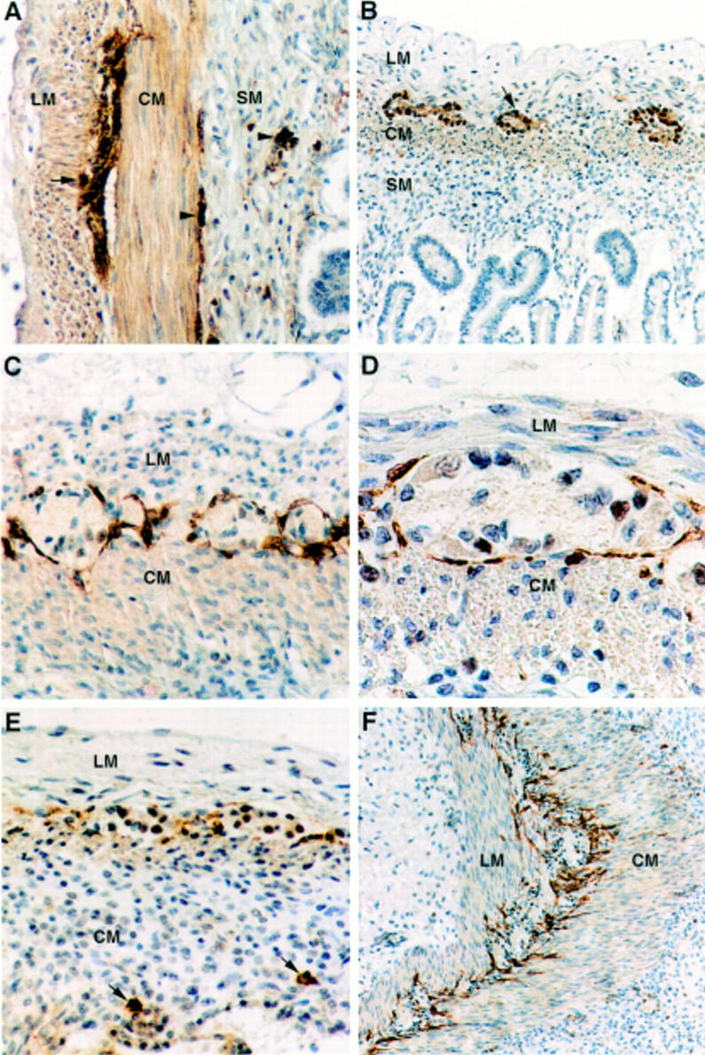
Figure 1 .

(A) NSE immunohistochemistry showing immunoreactive ganglion cells in the myenteric plexus (arrow) and in the inner and outer submucous plexuses (arrowheads) of the small bowel in an infant born at 26 weeks of gestation who died at the age of 17 days. Nerve fibres associated with the ganglia and in the circular muscle layer are also stained. Original magnification × 66. (B) NSE immunohistochemistry of the small bowel at 13 weeks of gestation displays the myenteric plexus (arrow), whereas no immunoreactive ganglion cells were present in the submucosa. Original magnification × 40. (C) From 17-18 weeks of gestation, the ICCs form a continuous layer along the myenteric plexus of the small bowel. This is illustrated in a case at 20 weeks of gestation using c-kit immunohistochemistry. Original magnification × 80. (D) c-kit immunoreactive cells are elongated in shape and have an ovoid nucleus as in this case at 20 weeks of gestation. The cell processes do not seem to penetrate into the ganglia. Original magnification × 200. (E) From 13 to 16 weeks of gestation, the layer of c-kit immunoreactive ICCs associated with the myenteric plexus of the small bowel appears to be disrupted. In the submucosa, isolated round c-kit immunoreactive cells, interpreted as mast cells, are seen (arrows). In this case the submucous plexus had not yet developed at 13 weeks of gestation, as shown in (B). Original magnification × 80. (F) From 17 weeks of gestation, the processes of the ICCs are often seen to penetrate into the circular muscle layer, whereas isolated ICCs in this layer are only rarely observed. Original magnification × 50. Abbreviations: LM, longitudinal muscle layer; CM, circular muscle layer; SM, submucosa.
