Abstract
OBJECTIVE—To evaluate the in vivo biocompatibility of three different devices following interventional closure of a patent ductus arteriosus (PDA) in an animal model. MATERIALS AND METHODS—A medical grade stainless steel coil (n = 8), a nickel/titanium coil (n = 10), and a polyvinylalcohol foam plug knitted on a titanium wire frame (n = 11) were used for interventional closure of PDA in a neonatal lamb model. The PDA had been maintained by repetitive angioplasty. Between one and 278 days after implantation the animals were killed and the ductal block removed. In addition to standard histology and scanning electron microscopy, immunohistochemical staining for biocompatibility screening was also undertaken. RESULTS—Electron microscopy revealed the growth of a cellular layer in a cobblestone pattern on the implant surfaces with blood contact, which was completed as early as five weeks after implantation of all devices. Immunohistochemical staining of these superficial cells showed an endothelial cell phenotype. After initial thrombus formation causing occlusion of the PDA after implantation there was ingrowth of fibromuscular cells resembling smooth muscle cells. Transformation of thrombotic material was completed within six weeks in the polyvinylalcohol plug and around the nickel/titanium coil, and within six months after implantation of the stainless steel coil. An implant related foreign body reaction was seen in only one of the stainless steel coil specimens and in two of the nickel/titanium coil specimens. CONCLUSION—After implantation, organisation of thrombotic material with ingrowth of fibromuscular cells was demonstrated in a material dependent time pattern. The time it took for endothelium to cover the implants was independent of the type of implant. Little or no inflammatory reaction of the surrounding tissue was seen nine months after implantation. Keywords: congenital heart disease; patent ductus arteriosus; catheter technique; biocompatibility
Full Text
The Full Text of this article is available as a PDF (182.5 KB).
Figure 1 .
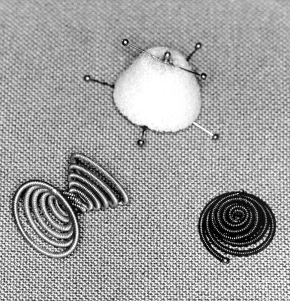
The implants. Top, foam polyvinylalcohol plug; bottom left, stainless steel coil; bottom right, nickel/titanium coil.
Figure 2 .
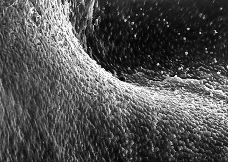
Cobblestone pattern of endothelial cells on the surface of the aortic orifice of a ductus arteriosus 112 days after interventional closure with a polyvinylalcohol plug (lamb 6). Electron microscopy × 600.
Figure 3 .
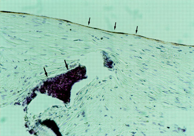
Positive immunohistochemical staining for von Willebrand factor in superficial endothelial cells (smaller arrows) covering the aortic orifice of a ductus arteriosus 117 days after implantation of a polyvinylalcohol plug. Larger arrows indicate foam material. × 800.
Figure 4 .
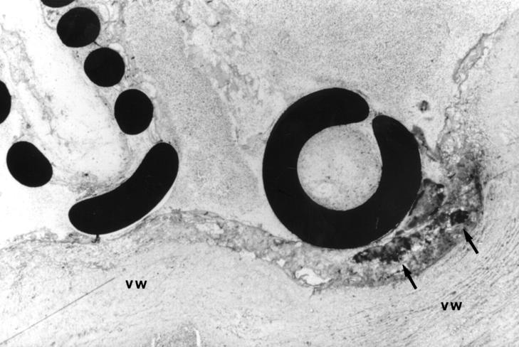
Central ductal portion filled with thrombotic material around coil loops in a lamb 107 days after stainless steel coil implantation. Methylmethacrylate embedded, ground section, toluidine blue stain. Black areas are the coil sections and the arrows indicate calcification. VW, vessel wall. × 200.
Figure 5 .
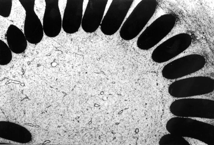
Central portion of an occluded ductus arteriosus in a lamb 32 days after nickel/titanium coil implantation, filled with fibromuscular cells. Methylmethacrylate embedded, ground section, toluidine blue stain. Black areas are the coil sections. × 200.
Selected References
These references are in PubMed. This may not be the complete list of references from this article.
- Abrams S. E., Walsh K. P., Diamond M. J., Clarkson M. J., Sibbons P. Radiofrequency thermal angioplasty maintains arterial duct patency. An experimental study. Circulation. 1994 Jul;90(1):442–448. doi: 10.1161/01.cir.90.1.442. [DOI] [PubMed] [Google Scholar]
- Burczak K., Gamian E., Kochman A. Long-term in vivo performance and biocompatibility of poly(vinyl alcohol) hydrogel macrocapsules for hybrid-type artificial pancreas. Biomaterials. 1996 Dec;17(24):2351–2356. doi: 10.1016/s0142-9612(96)00076-2. [DOI] [PubMed] [Google Scholar]
- Grabitz R. G., Freudenthal F., Sigler M., Le T. P., Boosfeld C., Handt S., von Bernuth G. Double-helix coil for occlusion of large patent ductus arteriosus: evaluation in a chronic lamb model. J Am Coll Cardiol. 1998 Mar 1;31(3):677–683. doi: 10.1016/s0735-1097(98)00025-4. [DOI] [PubMed] [Google Scholar]
- Grabitz R. G., Schräder R., Sigler M., Seghaye M. C., Dzionsko C., Handt S., Schneidt B., Von Bernuth G. Retrievable patent ductus arteriosus plug for interventional, transvenous occlusion of the patent ductus arteriosus. Evaluation in lambs and preliminary clinical results. Invest Radiol. 1997 Sep;32(9):523–528. doi: 10.1097/00004424-199709000-00004. [DOI] [PubMed] [Google Scholar]
- Lund G., Cragg A., Rysavy J., Castaneda F., Salomonowitz E., Vlodaver Z., Casteneda-Zuniga W., Amplatz K. Patency of the ductus arteriosus after balloon dilatation: an experimental study. Circulation. 1983 Sep;68(3):621–627. doi: 10.1161/01.cir.68.3.621. [DOI] [PubMed] [Google Scholar]
- Porstmann W., Wierny L., Warnke H. Der Verschluss des ductus arteriosus persistens ohne Thorakotomie. Radiol Diagn (Berl) 1968;9(2):168–169. [PubMed] [Google Scholar]
- Rao P. S., Wilson A. D., Sideris E. B., Chopra P. S. Transcatheter closure of patent ductus arteriosus with buttoned device: first successful clinical application in a child. Am Heart J. 1991 Jun;121(6 Pt 1):1799–1802. doi: 10.1016/0002-8703(91)90029-h. [DOI] [PubMed] [Google Scholar]
- Rigby M. L. Closure of the arterial duct: past, present, and future. Heart. 1996 Dec;76(6):461–462. doi: 10.1136/hrt.76.6.461. [DOI] [PMC free article] [PubMed] [Google Scholar]
- Rondelli G. Corrosion resistance tests on NiTi shape memory alloy. Biomaterials. 1996 Oct;17(20):2003–2008. doi: 10.1016/0142-9612(95)00352-5. [DOI] [PubMed] [Google Scholar]


