Full Text
The Full Text of this article is available as a PDF (91.7 KB).
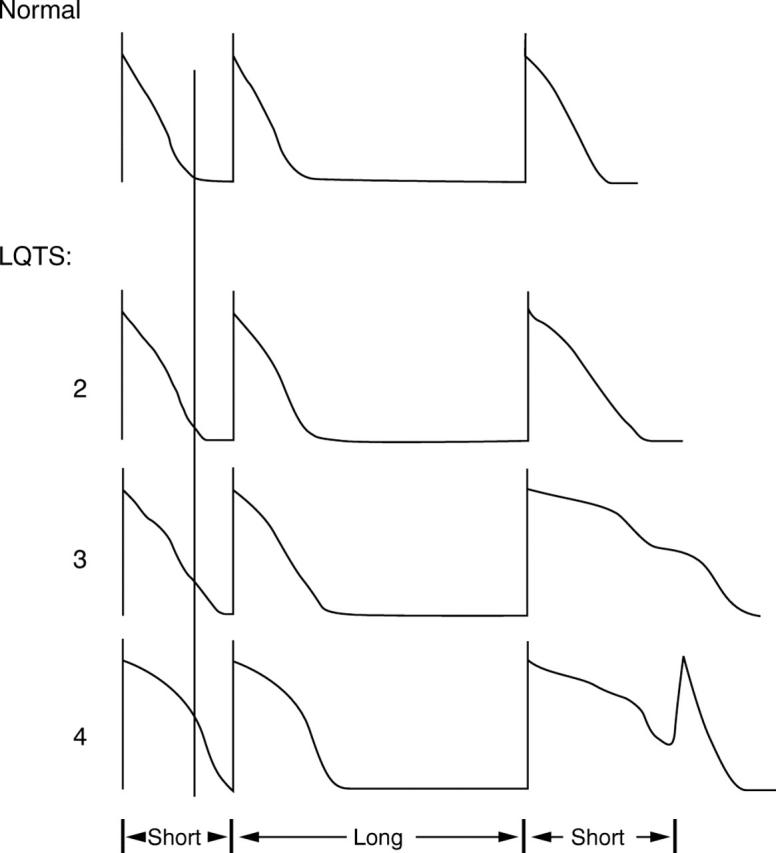
Figure 1 .

Schematic of cardiac action potentials before and during a pause. A normal response is shown in the top panel, and three action potentials (2, 3, and 4) in long QT syndrome (LQTS) below. Action potentials in LQTS are prolonged compared to normal, indicated by the vertical line. However, after the pause, the extent of action potential prolongation is exaggerated and highly heterogeneous, with an afterdepolarisation in the third panel and a triggered upstroke in the fourth. Under these conditions, the triggered beat may propagate to sites with short action potentials (for example, action potential 2) but not to sites with longer ones (for example, action potential 3), initiating the unstable re-entry that is thought to underlie torsades de pointes.17 A "short-long-short" series of cycles would appear on the surface ECG.


