

Abstract
Background: Children who survive very low birth weight (VLBW) without major disability have a high prevalence of learning difficulty, attention deficit, and dyspraxia.
Aims: To determine whether learning difficulty in children with VLBW is associated with structural brain abnormalities.
Methods: A total of 87 children (aged 15–16 years) with a history of VLBW (<1500 g) and eight age matched full term controls have been studied with detailed magnetic resonance brain scans. Volume measurements of the caudate nuclei and hippocampal formations were made.
Results: Scans in 42.5% of the children showed evidence of perinatal brain injury. There was no significant difference in IQ, dyspraxia, or attention deficit between children with qualitatively normal and abnormal scans. However, quantitative volumetric analysis showed that children with a low IQ had smaller volume measurements for the right caudate nucleus and left hippocampus, and a smaller hippocampal ratio (left volume:right volume) than those with normal IQ.
Conclusion: Data suggest that learning disorder, attention deficit, and dyspraxia in children who survive VLBW do not correlate with conventional markers of perinatal brain injury, and may be related to global brain growth and the development of key structures, such as the caudate nuclei and hippocampal formations.
Full Text
The Full Text of this article is available as a PDF (145.5 KB).
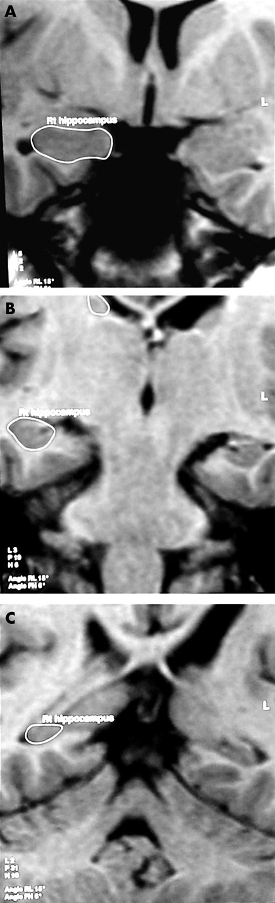
Figure 1 .
Coronal MR images showing the in-plane boundaries of the hippocampal formation: (A) head, (B) body, (C) posterior boundary at the level of the crus of the fornix.
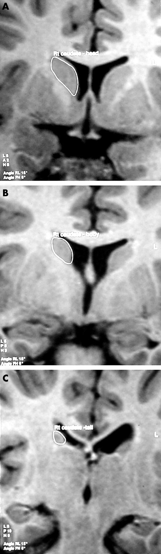
Figure 2 .
Coronal MR images showing the in-plane boundaries of the caudate nucleus: (A) head, (B) body, (C) tail.
Figure 3 .

Surface rendered 3-D reconstructions of the caudate nucleus and hippocampal formation generated from volume data on the Gyroview workstation.
Selected References
These references are in PubMed. This may not be the complete list of references from this article.
- Ajayi-Obe M., Saeed N., Cowan F. M., Rutherford M. A., Edwards A. D. Reduced development of cerebral cortex in extremely preterm infants. Lancet. 2000 Sep 30;356(9236):1162–1163. doi: 10.1016/s0140-6736(00)02761-6. [DOI] [PubMed] [Google Scholar]
- Bland J. M., Altman D. G. Statistical methods for assessing agreement between two methods of clinical measurement. Lancet. 1986 Feb 8;1(8476):307–310. [PubMed] [Google Scholar]
- Botting N., Powls A., Cooke R. W., Marlow N. Attention deficit hyperactivity disorders and other psychiatric outcomes in very low birthweight children at 12 years. J Child Psychol Psychiatry. 1997 Nov;38(8):931–941. doi: 10.1111/j.1469-7610.1997.tb01612.x. [DOI] [PubMed] [Google Scholar]
- Bremner J. D., Randall P., Scott T. M., Bronen R. A., Seibyl J. P., Southwick S. M., Delaney R. C., McCarthy G., Charney D. S., Innis R. B. MRI-based measurement of hippocampal volume in patients with combat-related posttraumatic stress disorder. Am J Psychiatry. 1995 Jul;152(7):973–981. doi: 10.1176/ajp.152.7.973. [DOI] [PMC free article] [PubMed] [Google Scholar]
- Castellanos F. X., Giedd J. N., Eckburg P., Marsh W. L., Vaituzis A. C., Kaysen D., Hamburger S. D., Rapoport J. L. Quantitative morphology of the caudate nucleus in attention deficit hyperactivity disorder. Am J Psychiatry. 1994 Dec;151(12):1791–1796. doi: 10.1176/ajp.151.12.1791. [DOI] [PubMed] [Google Scholar]
- Castellanos F. X., Giedd J. N., Marsh W. L., Hamburger S. D., Vaituzis A. C., Dickstein D. P., Sarfatti S. E., Vauss Y. C., Snell J. W., Lange N. Quantitative brain magnetic resonance imaging in attention-deficit hyperactivity disorder. Arch Gen Psychiatry. 1996 Jul;53(7):607–616. doi: 10.1001/archpsyc.1996.01830070053009. [DOI] [PubMed] [Google Scholar]
- Cooke R. W., Abernethy L. J. Cranial magnetic resonance imaging and school performance in very low birth weight infants in adolescence. Arch Dis Child Fetal Neonatal Ed. 1999 Sep;81(2):F116–F121. doi: 10.1136/fn.81.2.f116. [DOI] [PMC free article] [PubMed] [Google Scholar]
- Cooke R. W. Annual audit of three year outcome in very low birthweight infants. Arch Dis Child. 1993 Sep;69(3 Spec No):295–298. doi: 10.1136/adc.69.3_spec_no.295. [DOI] [PMC free article] [PubMed] [Google Scholar]
- Escobar G. J., Littenberg B., Petitti D. B. Outcome among surviving very low birthweight infants: a meta-analysis. Arch Dis Child. 1991 Feb;66(2):204–211. doi: 10.1136/adc.66.2.204. [DOI] [PMC free article] [PubMed] [Google Scholar]
- Filipek P. A., Semrud-Clikeman M., Steingard R. J., Renshaw P. F., Kennedy D. N., Biederman J. Volumetric MRI analysis comparing subjects having attention-deficit hyperactivity disorder with normal controls. Neurology. 1997 Mar;48(3):589–601. doi: 10.1212/wnl.48.3.589. [DOI] [PubMed] [Google Scholar]
- Giedd J. N., Castellanos F. X., Casey B. J., Kozuch P., King A. C., Hamburger S. D., Rapoport J. L. Quantitative morphology of the corpus callosum in attention deficit hyperactivity disorder. Am J Psychiatry. 1994 May;151(5):665–669. doi: 10.1176/ajp.151.5.665. [DOI] [PubMed] [Google Scholar]
- Giedd J. N., Snell J. W., Lange N., Rajapakse J. C., Casey B. J., Kozuch P. L., Vaituzis A. C., Vauss Y. C., Hamburger S. D., Kaysen D. Quantitative magnetic resonance imaging of human brain development: ages 4-18. Cereb Cortex. 1996 Jul-Aug;6(4):551–560. doi: 10.1093/cercor/6.4.551. [DOI] [PubMed] [Google Scholar]
- Hynd G. W., Hern K. L., Novey E. S., Eliopulos D., Marshall R., Gonzalez J. J., Voeller K. K. Attention deficit-hyperactivity disorder and asymmetry of the caudate nucleus. J Child Neurol. 1993 Oct;8(4):339–347. doi: 10.1177/088307389300800409. [DOI] [PubMed] [Google Scholar]
- Hynd G. W., Semrud-Clikeman M., Lorys A. R., Novey E. S., Eliopulos D. Brain morphology in developmental dyslexia and attention deficit disorder/hyperactivity. Arch Neurol. 1990 Aug;47(8):919–926. doi: 10.1001/archneur.1990.00530080107018. [DOI] [PubMed] [Google Scholar]
- Hynd G. W., Semrud-Clikeman M., Lorys A. R., Novey E. S., Eliopulos D., Lyytinen H. Corpus callosum morphology in attention deficit-hyperactivity disorder: morphometric analysis of MRI. J Learn Disabil. 1991 Mar;24(3):141–146. doi: 10.1177/002221949102400302. [DOI] [PubMed] [Google Scholar]
- Jack C. R., Jr, Bentley M. D., Twomey C. K., Zinsmeister A. R. MR imaging-based volume measurements of the hippocampal formation and anterior temporal lobe: validation studies. Radiology. 1990 Jul;176(1):205–209. doi: 10.1148/radiology.176.1.2353093. [DOI] [PubMed] [Google Scholar]
- Jack C. R., Jr MRI-based hippocampal volume measurements in epilepsy. Epilepsia. 1994;35 (Suppl 6):S21–S29. doi: 10.1111/j.1528-1157.1994.tb05986.x. [DOI] [PubMed] [Google Scholar]
- Marlow N., Roberts B. L., Cooke R. W. Laterality and prematurity. Arch Dis Child. 1989 Dec;64(12):1713–1716. doi: 10.1136/adc.64.12.1713. [DOI] [PMC free article] [PubMed] [Google Scholar]
- Marlow N., Roberts L., Cooke R. Outcome at 8 years for children with birth weights of 1250 g or less. Arch Dis Child. 1993 Mar;68(3 Spec No):286–290. doi: 10.1136/adc.68.3_spec_no.286. [DOI] [PMC free article] [PubMed] [Google Scholar]
- Mataró M., Garcia-Sánchez C., Junqué C., Estévez-González A., Pujol J. Magnetic resonance imaging measurement of the caudate nucleus in adolescents with attention-deficit hyperactivity disorder and its relationship with neuropsychological and behavioral measures. Arch Neurol. 1997 Aug;54(8):963–968. doi: 10.1001/archneur.1997.00550200027006. [DOI] [PubMed] [Google Scholar]
- Mercuri E., Faundez J. C., Roberts I., Flora S., Bouza H., Cowan F., Pennock J., Bydder G., Dubowitz L. Neurological 'soft' signs may identify children with sickle cell disease who are at risk for stroke. Eur J Pediatr. 1995 Feb;154(2):150–156. [PubMed] [Google Scholar]
- Obenaus A., Yong-Hing C. J., Tong K. A., Sarty G. E. A reliable method for measurement and normalization of pediatric hippocampal volumes. Pediatr Res. 2001 Jul;50(1):124–132. doi: 10.1203/00006450-200107000-00022. [DOI] [PubMed] [Google Scholar]
- Olsén P., Päkkö E., Vainionpä L., Pyhtinen J., Järvelin M. R. Magnetic resonance imaging of periventricular leukomalacia and its clinical correlation in children. Ann Neurol. 1997 Jun;41(6):754–761. doi: 10.1002/ana.410410611. [DOI] [PubMed] [Google Scholar]
- Peterson B. S., Vohr B., Staib L. H., Cannistraci C. J., Dolberg A., Schneider K. C., Katz K. H., Westerveld M., Sparrow S., Anderson A. W. Regional brain volume abnormalities and long-term cognitive outcome in preterm infants. JAMA. 2000 Oct 18;284(15):1939–1947. doi: 10.1001/jama.284.15.1939. [DOI] [PubMed] [Google Scholar]
- Powls A., Botting N., Cooke R. W., Marlow N. Motor impairment in children 12 to 13 years old with a birthweight of less than 1250 g. Arch Dis Child Fetal Neonatal Ed. 1995 Sep;73(2):F62–F66. doi: 10.1136/fn.73.2.f62. [DOI] [PMC free article] [PubMed] [Google Scholar]
- Powls A., Botting N., Cooke R. W., Pilling D., Marlow N. Growth impairment in very low birthweight children at 12 years: correlation with perinatal and outcome variables. Arch Dis Child Fetal Neonatal Ed. 1996 Nov;75(3):F152–F157. doi: 10.1136/fn.75.3.f152. [DOI] [PMC free article] [PubMed] [Google Scholar]
- Santosh P. J. Neuroimaging in child and adolescent psychiatric disorders. Arch Dis Child. 2000 May;82(5):412–419. doi: 10.1136/adc.82.5.412. [DOI] [PMC free article] [PubMed] [Google Scholar]
- Szabó C. A., Wyllie E., Siavalas E. L., Najm I., Ruggieri P., Kotagal P., Lüders H. Hippocampal volumetry in children 6 years or younger: assessment of children with and without complex febrile seizures. Epilepsy Res. 1999 Jan;33(1):1–9. doi: 10.1016/s0920-1211(98)00068-0. [DOI] [PubMed] [Google Scholar]
- Watson C., Andermann F., Gloor P., Jones-Gotman M., Peters T., Evans A., Olivier A., Melanson D., Leroux G. Anatomic basis of amygdaloid and hippocampal volume measurement by magnetic resonance imaging. Neurology. 1992 Sep;42(9):1743–1750. doi: 10.1212/wnl.42.9.1743. [DOI] [PubMed] [Google Scholar]
- de Vries L. S., Eken P., Groenendaal F., van Haastert I. C., Meiners L. C. Correlation between the degree of periventricular leukomalacia diagnosed using cranial ultrasound and MRI later in infancy in children with cerebral palsy. Neuropediatrics. 1993 Oct;24(5):263–268. doi: 10.1055/s-2008-1071554. [DOI] [PubMed] [Google Scholar]