

Full Text
The Full Text of this article is available as a PDF (119.3 KB).
Figure 1 .
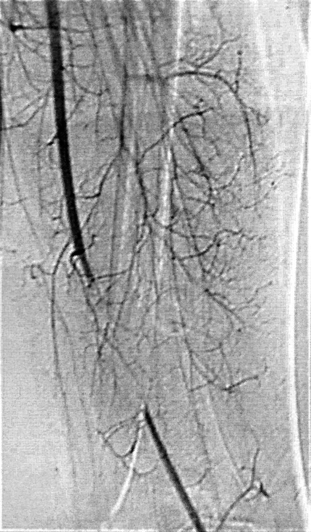
Arteriographic imaging showing superficial femoral artery interruption and collateral circulation.
Figure 2 .

(A) The ischaemic area at the heel, shown by arrows, 48 hours after fasciotomy. (B) Evolution of the necrotic area at the heel (pre-escharotomy), with good revascularisation of pre-existing ischaemic area after one week NGF treatment. Ischaemic lesions of the toes persist unchanged in size, and formation of bullous lesions can be observed.