

Abstract
Background: Previous studies have used the dynamic susceptibility contrast enhanced (DSCE) magnetic resonance (MR) imaging technique to measure cerebral perfusion in adults.
Objective: To assess the feasibility of the technique in a heterogeneous cohort of sick human infants and identify cerebral perfusion abnormalities.
Methods: Perfusion measurements were made by characterising the changing concentration of an injected bolus of contrast agent using a series of MR images acquired during the first pass of the contrast bolus. Qualitative values of relative cerebral blood flow (rCBF) were then calculated from these data on a pixel by pixel basis to generate parametric maps of perfusion.
Results: Images of perfusion were successfully calculated from 12 out of 27 neonates and infants, all with established cerebral pathology. Normal vascular anatomical structures such as the circle of Willis were identified within all calculated images. Values of rCBF were generally larger in grey matter than in white matter. In several patients, perfusion abnormalities resulted in structural abnormalities which were detected in conventional MR imaging at follow up. The acquisition of perfusion data was most difficult when the least mature brains were examined because of motion artefacts and a smaller head size with a lower level of rCBF than adults.
Conclusions: This preliminary study shows that: (a) maps of rCBF can be acquired from neonates and infants; (b) characterisation of the bolus passage becomes progressively easier as the brain matures; (c) early abnormalities in cerebral perfusion may have negative prognostic implications; (d) the main difficulty when using the DSCE technique to study neonates relates to image artefacts resulting from bulk head motion.
Full Text
The Full Text of this article is available as a PDF (806.9 KB).
Figure 1 .
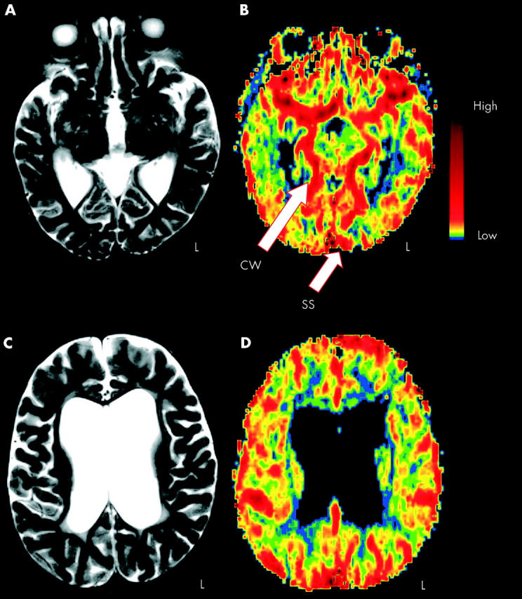
Magnetic resonance (MR) structural-functional comparison. Conventional T2 weighted images (A and C) versus MR perfusion images (B and D); axial sections obtained at two levels of the brain. Functional data are displayed using a non-linear colour scale. CW, Circle of Willis; SS, sagittal sinus; L, left. See text for further details.
Figure 2 .

Magnetic resonance (MR) structural-functional comparison in hypoxic ischaemic encephalopathy. Conventional T2 weighted images (A and C) versus MR perfusion images (B and D); axial sections obtained at two levels of the brain. Colour conventions as in fig 1. Arrows indicate regions of hypoperfusion in the occipital cortex at the age of 13 days (B) corresponding to cystic lesions in the conventional MR at the age of 10 weeks (C). A–C were acquired from a patient with hypoxic ischaemic encephalopathy, whereas D was obtained from a different patient with convulsions. See text for further details.
Selected References
These references are in PubMed. This may not be the complete list of references from this article.
- Barkovich A. J., Latal-Hajnal B., Partridge J. C., Sola A., Ferriero D. M. MR contrast enhancement of the normal neonatal brain. AJNR Am J Neuroradiol. 1997 Oct;18(9):1713–1717. [PMC free article] [PubMed] [Google Scholar]
- Calamante F., Thomas D. L., Pell G. S., Wiersma J., Turner R. Measuring cerebral blood flow using magnetic resonance imaging techniques. J Cereb Blood Flow Metab. 1999 Jul;19(7):701–735. doi: 10.1097/00004647-199907000-00001. [DOI] [PubMed] [Google Scholar]
- Gore J. C., Majumdar S. Measurement of tissue blood flow using intravascular relaxation agents and magnetic resonance imaging. Magn Reson Med. 1990 May;14(2):242–248. doi: 10.1002/mrm.1910140210. [DOI] [PubMed] [Google Scholar]
- Greisen G., Børch K. White matter injury in the preterm neonate: the role of perfusion. Dev Neurosci. 2001;23(3):209–212. doi: 10.1159/000046145. [DOI] [PubMed] [Google Scholar]
- Greisen G., Johansen K., Ellison P. H., Fredriksen P. S., Mali J., Friis-Hansen B. Cerebral blood flow in the newborn infant: comparison of Doppler ultrasound and 133xenon clearance. J Pediatr. 1984 Mar;104(3):411–418. doi: 10.1016/s0022-3476(84)81108-7. [DOI] [PubMed] [Google Scholar]
- Reith W., Heiland S., Erb G., Benner T., Forsting M., Sartor K. Dynamic contrast-enhanced T2*-weighted MRI in patients with cerebrovascular disease. Neuroradiology. 1997 Apr;39(4):250–257. doi: 10.1007/s002340050403. [DOI] [PubMed] [Google Scholar]
- Rempp K. A., Brix G., Wenz F., Becker C. R., Gückel F., Lorenz W. J. Quantification of regional cerebral blood flow and volume with dynamic susceptibility contrast-enhanced MR imaging. Radiology. 1994 Dec;193(3):637–641. doi: 10.1148/radiology.193.3.7972800. [DOI] [PubMed] [Google Scholar]
- Tokumaru A. M., Barkovich A. J., O'uchi T., Matsuo T., Kusano S. The evolution of cerebral blood flow in the developing brain: evaluation with iodine-123 iodoamphetamine SPECT and correlation with MR imaging. AJNR Am J Neuroradiol. 1999 May;20(5):845–852. [PMC free article] [PubMed] [Google Scholar]
- Volpe J. J., Herscovitch P., Perlman J. M., Raichle M. E. Positron emission tomography in the newborn: extensive impairment of regional cerebral blood flow with intraventricular hemorrhage and hemorrhagic intracerebral involvement. Pediatrics. 1983 Nov;72(5):589–601. [PubMed] [Google Scholar]