



Abstract
Aberrant subclavian artery (arteria lusoria) is a rare congenital anomaly that usually does not produce symptoms. Symptomatic patients require surgical intervention. The operative approach to correct this condition has been controversial. Herein, we describe surgical approaches to the aberrant right subclavian artery.
From 2000 through 2004, 3 children and 1 adult with aberrant right subclavian artery underwent operation. Our surgical approach varied according to the age of the patient. A muscle-sparing right thoracotomy was used in the pediatric patients, and a supraclavicular approach was used in the adult. Patients were treated successfully by division of the aberrant right subclavian artery and translocation to the right common carotid artery, without graft interposition. There was no operative or late morbidity or death. Symptoms were completely relieved in all patients.
Although an extrathoracic approach is applicable and reliable for adult patients, we believe that adequate exposure for the described procedure is best accomplished through a right thoracotomy in pediatric patients. This approach enables optimal mobilization of the distal right subclavian artery without leaving a long stump and enables direct anastomosis to the ipsilateral carotid artery.
Key words: Adult; aorta, thoracic/abnormalities, child, deglutition disorders/etiology, esophageal stenosis/etiology, subclavian artery/abnormalities/surgery, respiratory insufficiency/etiology, vascular surgical procedures/methods
Aberrant right subclavian artery (RSA) is a rare congenital anomaly that usually does not produce symptoms. Symptomatic patients require surgical intervention. When surgical treatment is indicated, consideration should be given to anatomic reestablishment of orthograde flow into the right subclavian artery, which avoids sacrificing direct blood flow to the right arm and the possible complications thereof. Many surgical approaches, such as median sternotomy, left and right thoracotomies, and supraclavicular incision, have been used to accomplish this objective. Nevertheless, the surgical approach to repair this condition has been controversial. We present the cases of 3 children and 1 adult who were successfully treated, through different surgical approaches, by division and translocation of the aberrant right subclavian artery to the right common carotid artery, without graft interposition.
Patients and Methods
From 2000 through 2004, 3 children and 1 adult underwent surgery for aberrant RSA at our institution. Patients' characteristics are summarized in Table I. All patients presented with a history of difficulty in swallowing solid food. In addition, respiratory problems were seen in 2 of the 3 children. Barium-contrast examinations of the esophagus showed indentations at the upper thoracic esophageal level in all patients (Fig. 1A). Other examinations, such as digital subtraction angiography (Fig. 2), contrast-enhanced magnetic resonance angiography, and computed tomography, confirmed the presence of an aberrant RSA originating from the aortic arch below the left subclavian artery. There was no brachiocephalic trunk, and the 1st cephalic artery was the right common carotid, followed by the left common carotid artery and the left subclavian artery (Fig. 2).
TABLE I. Characteristics and Findings in the 4 Patients


Fig. 1 A) Preoperative esophagram shows marked posterior compression (arrow) on the proximal esophagus in patient 2. B) Postoperative esophagram shows release of compression on the proximal esophagus in the same patient.

Fig. 2 Digital subtraction angiogram of the thoracic aorta shows aberrant right subclavian artery (arrow) in patient 4.
Surgical Technique
The bilateral radial arteries were monitored. A thoracic tube (24F–32F) was inserted into the esophagus for easy exploration.
The adult patient (40 years old) underwent surgery via a single right supraclavicular approach. Injury to the brachial plexus and the right recurrent laryngeal nerve was avoided by careful identification of these structures. The subclavian artery was divided approximately 1 cm distal to the aortic arch. The proximal portion was oversewn, and the distal portion was anastomosed to the carotid artery.
In the pediatric patients, the chest was entered through a right anterolateral thoracotomy in the 4th intercostal space with a muscle-sparing technique. The aberrant right subclavian artery arose from the posterior left subclavian artery at the distal aortic arch (Fig. 3). In all cases, the vagus and recurrent laryngeal nerves were identified and preserved. The anomalous vessel was dissected free from its retroesophageal position and was subsequently divided at its origin at the aortic arch. After division, the proximal end was retracted to the anterior of the esophagus and anastomosed to the right carotid artery.

Fig. 3 Intraoperative view of aberrant right subclavian artery (clamped) in patient 3.
Results
Recovery was uneventful in all patients. We did not observe any evidence of neural damage during the postoperative period. All patients were asymptomatic, and digital subtraction or magnetic resonance angiography (Fig. 4) performed at 1 week to 2 months after surgery revealed a widely patent carotid–subclavian artery anastomosis. The indentation in the esophagus disappeared in follow-up barium-contrast examination in all patients (Fig. 1B).
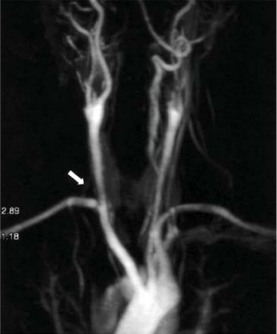
Fig. 4 Postoperative magnetic resonance angiogram shows translocation of the aberrant right subclavian artery to the right common carotid artery (arrow) in patient 4.
Discussion
Aberrant subclavian artery, also called arteria lusoria, was first described by Bayford in 1794 in a 62-year-old woman who died after years of dysphagia.1 The most common form of aortic arch anomaly, aberrant subclavian artery, has an estimated occurrence rate of 0.5%.2 It results from regression of the right 4th aortic arch between the carotid and subclavian arteries, rather than distal to the subclavian artery.3,4 The right subclavian artery usually persists as a branch from the descending aorta coursing posterior to the esophagus, but it may pass between the esophagus and the trachea, or even anterior to the trachea.
The most common symptoms include dysphagia, cough, stridor, and thoracic pain; these are usually associated with evident compression of the adjacent structure. On the basis of autopsy studies and retrospective analyses of patients' symptoms during life, we may conclude that most patients remain symptom-free during their lives.5 Adults typically present with symptoms of dysphagia; infants more often present with respiratory symptoms. Moreover, the increased frequency of pulmonary infection seen in infants who have this anomaly is not seen in older age groups. Respiratory problems in infancy are thought to be due to the absence of tracheal rigidity, in combination with dysphagia and aspiration of food particles. It is not clear why dysphagia may develop in the adult patient. Aneurysmal dilatation,6 increased rigidity of the esophagus itself or of the vascular wall, and fibrous transformation of the paratracheal and esophageal tissues—together with the narrow topologic vicinity of the proximal aortic branches, the age-related atheromatous process, aortic elongation, and the combination of an aberrant artery and a truncus bicaroticus7,8—may all play roles in the development of dysphagia.
Barium-contrast examination of the esophagus, showing a characteristic diagonal compression defect at the level of the 3rd and 4th vertebrae, is an excellent tool for diagnosing this condition. Computed tomographic scanning, angiography, or both, usually confirm the diagnosis. New imaging techniques such as magnetic resonance imaging may contribute to better visualization, especially when an aneurysm is present in the proximal part of the artery.
Surgical intervention is indicated for all patients who have symptomatic or aneurysmal aberrant RSA. In 1946, Gross9 performed the 1st operation to repair this anomaly. At first, treatment for aberrant RSA consisted of ligation of the vessel. Yet the goal of operative repair is to relieve the symptoms caused by the aberrant artery and to restore circulation. Because of previous reports of ischemia and noteworthy subclavian steal in open surgical correction of dysphagia lusoria,10 the re-establishment of flow at initial surgery, by either reimplantation or bypass, has been recommended.2 Although there are few reports of success with endovascular occlusion of aberrant RSA,11 that option appears to be valuable in treating elderly patients with comorbidities that make them unsuitable for major surgery.
Many reports have emphasized the importance of restoring pulsatile blood flow to the right subclavian artery.12–14 This is accomplished by anastomosis of the divided subclavian artery to the ascending aorta or the right common carotid artery (Fig. 5) either directly or by use of a short interposition graft. Both our experience and our review of the English-language medical literature indicate that subclavian carotid transposition is an ideal technique for reconstruction of this artery, with excellent long-term patency.15

Fig. 5 Schematic drawing shows path of the aberrant right subclavian artery (RSA) and surgical correction.
There is no standard surgical approach to the repair of this anomaly. Right and left thoracotomies, cervical incision, median sternotomy, and combinations of these approaches have been used. In the largest published series of treated aberrant RSA, Kieffer and colleagues2 included 33 cases with basically different therapeutic strategies. Conclusively, the authors proposed classifying patients into 4 aberrant RSA groups depending on their anatomical characteristics:
Group 1 – Patients with dysphagia caused by nonaneurysmal aberrant RSA;
Group 2 – Patients with symptomatic occlusive disease of nonaneurysmal aberrant RSA;
Group 3 – Patients with aneurysmal aberrant RSA without aortic lesions, with or without esophageal compression or arterial thromboembolism;
Group 4 – Patients with an aortic (usually aneurysmal) lesion involving the origin of the aberrant RSA, with or without aberrant RSA aneurysm.2
We think that the choice of approach depends on the presence or absence of aneurysmal formation and on the age of the patient. When there is an aberrant RSA aneurysm, thoracotomy or median sternotomy is needed. Many authors have suggested that there is no need for thoracotomy or extensive dissection, instead advocating a right supraclavicular approach through a single incision to achieve the surgical goals of ligation, reimplantation, and esophageal dilation.16–19 Indeed, adult patients who have an aberrant subclavian artery uncomplicated by aneurysm or aortic involvement can undergo successful operative repair via a single-incision supraclavicular approach. In our literature review, we found that this technique was generally used in adult patients. In children, the supraclavicular approach has the disadvantages of less favorable exposure of the vessel and more difficult control of hemorrhaging. In addition, a possible consequence of less favorable operative exposure is a persistent long arterial stump behind the esophagus, which might cause ongoing symptoms, embolization, or aneurysmal dilatation. There are, however, no reported data to support these conjectures.
Van Son and coworkers13 found that the aberrant RSA originates from the posteromedial side of the distal aortic arch and that a surgical approach through a right thoracotomy enables mobilization of the vessel, division at its origin without leaving a long stump, and connection to the ascending aorta or right common carotid artery.
Keiffer and colleagues have emphasized the anatomic variations that are associated with aberrant RSA: these include abnormal origin of the right vertebral artery from the aorta or from the right common carotid artery, the presence of a common carotid trunk, a right-sided thoracic duct, and a nonrecurrent laryngeal nerve.2 When the laryngeal nerve is nonrecurrent, it arises from the vagus nerve in the neck and directly innervates the larynx. Although this anomaly is of less importance for surgical treatment of aberrant RSA than are the others, it is important to recognize in patients who may require a carotid artery or thyroid procedure. Our patients did not experience any neural palsy during the postoperative period. To achieve this, the surgeon must identify and protect the vagus and recurrent laryngeal nerves while performing vascular exploration. In the posterior mediastinal exploration, dissection close to the vessel may decrease the risk of neural damage. We think that right thoracotomy decreases the risk of laryngeal nerve damage in nonaneurysmal patients—especially the risk of damage to the left recurrent nerve, which courses close to the origin of the aberrant RSA.
We believe that the best mediastinal exposure for the described procedure is accomplished through a right thoracotomy, especially in pediatric patients. This approach enables optimal mobilization to the distal right subclavian artery and direct anastomosis to the ipsilateral carotid artery, without graft interposition. Several other approaches, mainly a left thoracotomy in combination with a cervical approach or an extrathoracic approach, have the disadvantages of less favorable exposure of the vessel and more difficult control of hemorrhaging, as well as the need to position the patient twice for 2 incisions.
Footnotes
Address for reprints: Yuksel Atay, MD, Department of Cardiovascular Surgery, Ege University Hospital, Bornova, 35100 Izmir, Turkey. E-mail: yuksel.atay@ege.edu.tr
References
- 1.Bayford D. An account of a singular case of obstructed deglutition. Memoirs Med Soc London 1794;2:275–86.
- 2.Kieffer E, Bahnini A, Koskas F. Aberrant subclavian artery: surgical treatment in thirty-three adult patients. J Vasc Surg 1994;19:100–11. [DOI] [PubMed]
- 3.Bull PG, Denck H. Aberrant right subclavian artery. Eur J Vasc Surg 1994;8:757–60. [DOI] [PubMed]
- 4.Freed K, Low VH. The aberrant subclavian artery. AJR Am J Roentgenol 1997;168:481–4. [DOI] [PubMed]
- 5.Molz G, Burri B. Aberrant subclavian artery (arteria lusoria): sex differences in the prevalence of various forms of the malformation. Evaluation of 1378 observations. Virchows Arch A Pathol Anat Histol 1978;380:303–15. [DOI] [PubMed]
- 6.Brown DL, Chapman WC, Edwards WH, Coltharp WH, Stoney WS. Dysphagia lusoria: aberrant right subclavian artery with a Kommerell's diverticulum. Am Surg 1993;59:582–6. [PubMed]
- 7.Klinkhamer AC. Het vaststellen van aberrante arterien in het mediastinum superius door middel van oesophagogram [thesis]. Leyden; 1962. p. 1–187.
- 8.Klinkhamer AC. Aberrant right subclavian artery. Clinical and roentgenologic aspects. Am J Roentgenol Radium Ther Nucl Med 1966;97:438–46. [DOI] [PubMed]
- 9.Gross RE. Surgical treatment for dysphagia lusoria. Ann Surg 1946;124:532–4. [PubMed]
- 10.Mok CK, Cheung KL, Kong SM, Ong GB. Translocating the aberrant right subclavian artery in dysphagia lusoria. Br J Surg 1979;66:113–6. [DOI] [PubMed]
- 11.Hausegger KA, Oberwalder P, Tiesenhausen K, Tauss J, Stanger O, Schedlbauer P, et al. Intentional left subclavian artery occlusion by thoracic aortic stent-grafts without surgical transposition. J Endovasc Ther 2001;8:472–6. [DOI] [PubMed]
- 12.Verkroost MW, Hamerlijnck RP, Vermeulen FE. Surgical management of aneurysms at the origin of an aberrant right subclavian artery. J Thorac Cardiovasc Surg 1994;107:1469–71. [PubMed]
- 13.van Son JA, Mierzwa M, Mohr FW. Resection of atherosclerotic aneurysm at origin of aberrant right subclavian artery. Eur J Cardiothorac Surg 1999;16:576–9. [DOI] [PubMed]
- 14.Stone WM, Brewster DC, Moncure AC, Franklin DP, Cambria RP, Abbott WM. Aberrant right subclavian artery: varied presentations and management options. J Vasc Surg 1990;11:812–7. [PubMed]
- 15.Cina CS, Arena GO, Bruin G, Clase CM. Kommerell's diverticulum and aneurysmal right-sided aortic arch: a case report and review of the literature. J Vasc Surg 2000;32:1208–14. [DOI] [PubMed]
- 16.Orvald TO, Scheerer R, Jude JR. A single cervical approach to aberrant right subclavian artery. Surgery 1972;71:227–30. [PubMed]
- 17.Valentine RJ, Carter DJ, Clagett GP. A modiied extrathoracic approach to the treatment of dysphagia lusoria. J Vasc Surg 1987;5:498–500. [PubMed]
- 18.Taylor M, Harris KA, Casson AG, DeRose G, Jamieson WG. Dysphagia lusoria: extrathoracic surgical management. Can J Surg 1996;39:48–52. [PMC free article] [PubMed]
- 19.Carrizo GJ, Marjani MA. Dysphagia lusoria caused by an aberrant right subclavian artery. Tex Heart Inst J 2004;31:168–71. [PMC free article] [PubMed]