Full Text
The Full Text of this article is available as a PDF (469.4 KB).
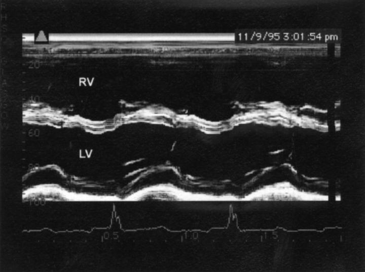
Figure 1 .
M mode study from a patient with a large secundum atrial septal defect. There is marked increase in the right ventricle diastolic dimension, and paradoxical septal motion with the septum moving in parallel with the posterior left ventricular wall. RV, right ventricle; LV, left ventricle.
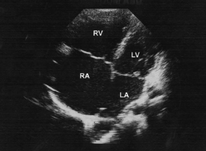
Figure 2 .
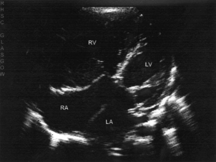
Parasternal four chamber view in a patient with a large secundum atrial septal defect. There is enlargement of the right ventricle and a large gap in the central part of the atrial septum. RA, right atrium; LA, left atrium; others as before.
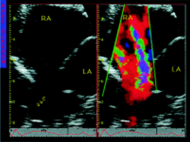
Figure 3 .
Cross sectional and colour images of a high sinus venosus atrial septal defect. The scanning plane has been tilted upwards to show the entry of the superior vena cava, which is shown to override the atrial septum. An anomalous pulmonary vein is not shown. SVC, superior vena cava; others as before.
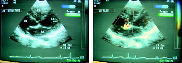
Figure 4 .
Cross sectional and colour images from a woman with an atrial septal aneurysm. (A) The aneurysm bulges into the right atrium and there are areas of echo dropout which might suggest a defect. (B) Colour is necessary to determine whether there is a defect and identify its site.
Figure 5 .
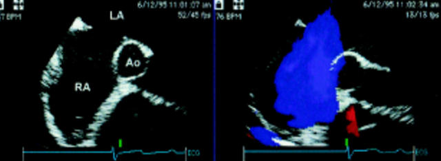
Transoesophageal echocardiography images in a patient with a large secundum atrial septal defect showing the defect and flow from the left to right atrium. The defect reaches to near the aorta and only a small edge of atrial septum is seen next to it at this margin. Ao, aortic root; others as before.
Figure 6 .
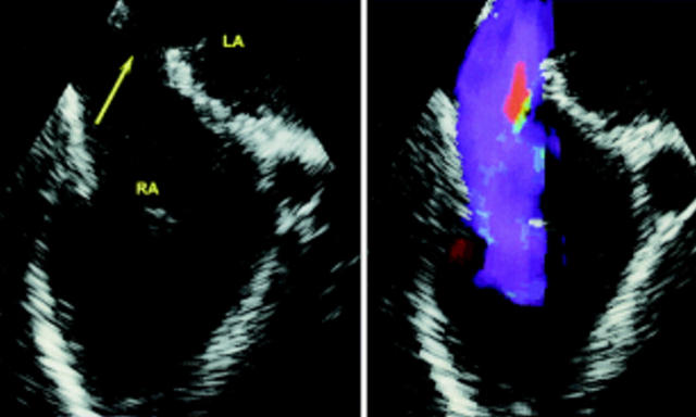
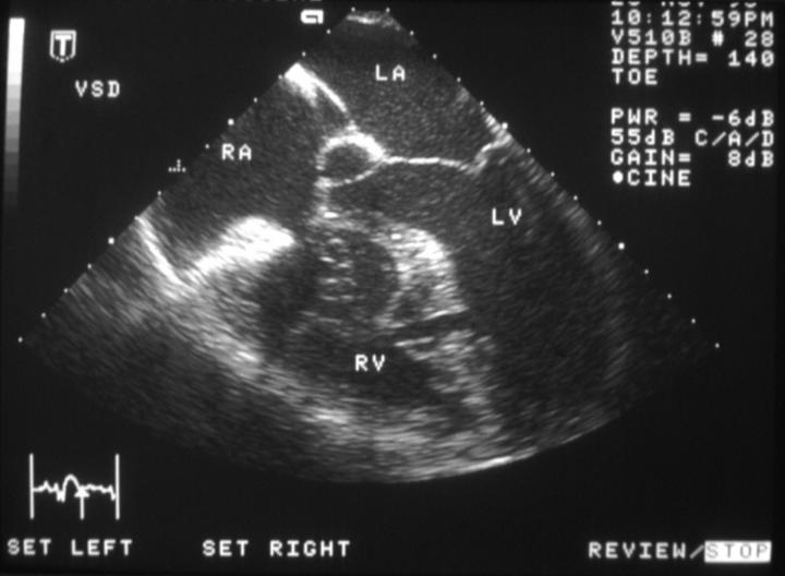
Transoesophageal echocardiography images in a patient with a sinus venosus atrial septal defect showing the defect (arrow) and flow from the left (LA) to right (RA) atrium. The defect is shown in the upper and posterior part of the septum at the entrance of the superior vena cava.
Figure 7 .
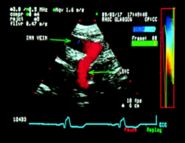
Left parasaggital view in a patient with partial anomalous pulmonary venous drainage of the left upper pulmonary vein to the innominate. The pulmonary vein enters a left ascending vein (LSVC) which joins the innominate vein (Inn vein). Colour shows the flow is upwards to the innominate.
Figure 8 .
Parasternal four chamber view in a patient with an ostium primum atrial septal defect. The atrioventricular valves are attached to the edge of the ventricular septum at the same level and there is a large gap in the septum primum between the ventricular and the atrial septae.
Figure 9 .
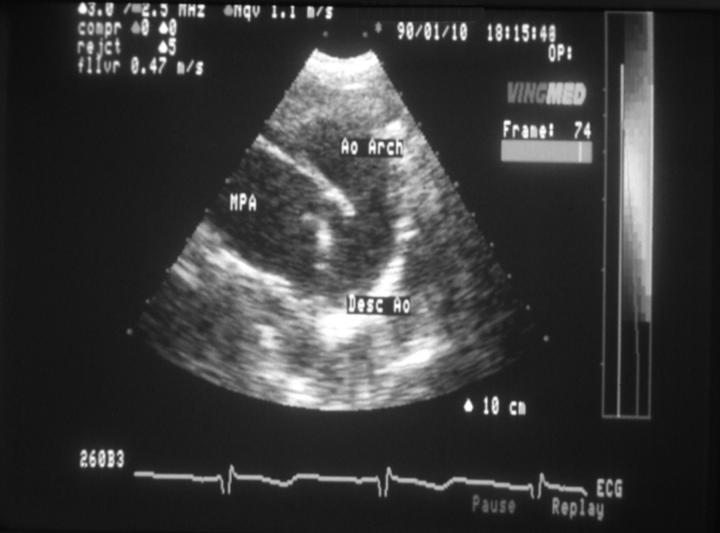
Left parasaggital view of the arterial duct in a teenage girl. The duct is shown as a continuation of the main pulmonary artery (MPA) into the descending aorta (Desc Ao). Ao Arch, aortic arch; others as before.
Figure 10 .
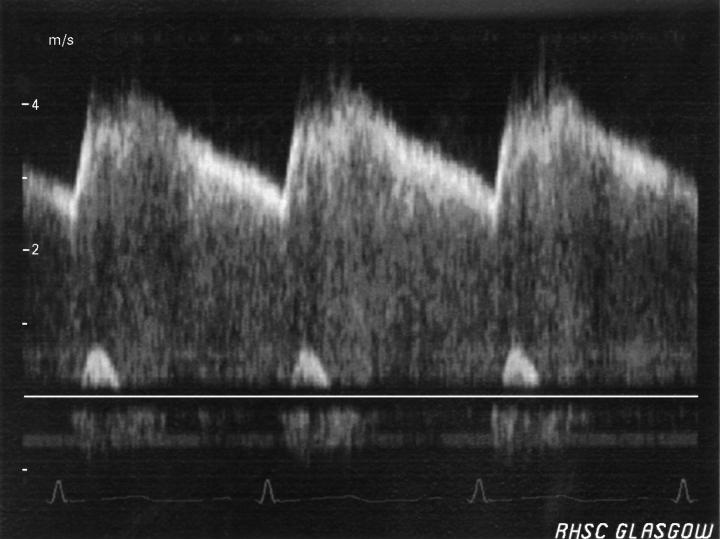
Spectral Doppler signal of ductal flow in ductus arteriosus with low pulmonary artery pressure. The signal is a continuous one reaching its maximum velocity (4.3 m/s) in mid-systole and then falls off until just after the R wave.
Figure 11 .
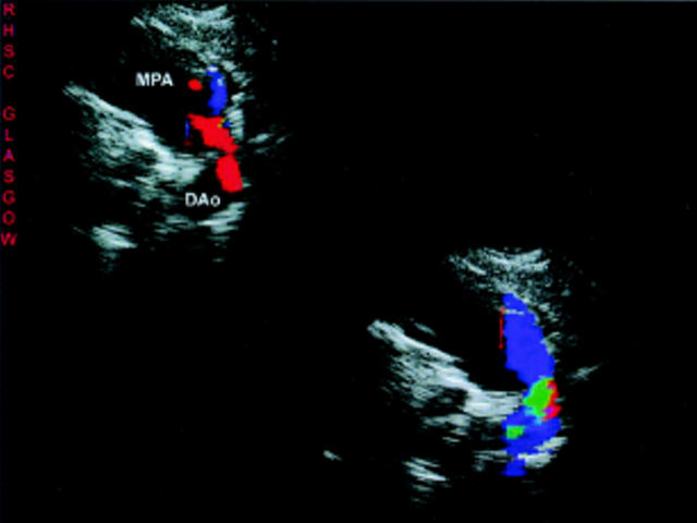
Left parasternal views in a woman with Eisenmenger's syndrome and patent ductus arteriosus. The duct is imaged as a continuation of the main pulmonary artery (MPA) into the descending aorta (DAo). Flow is bidirectional, from the pulmonary artery to aorta in systole (top) and aorta to pulmonary artery in diastole (bottom).
Figure 12 .
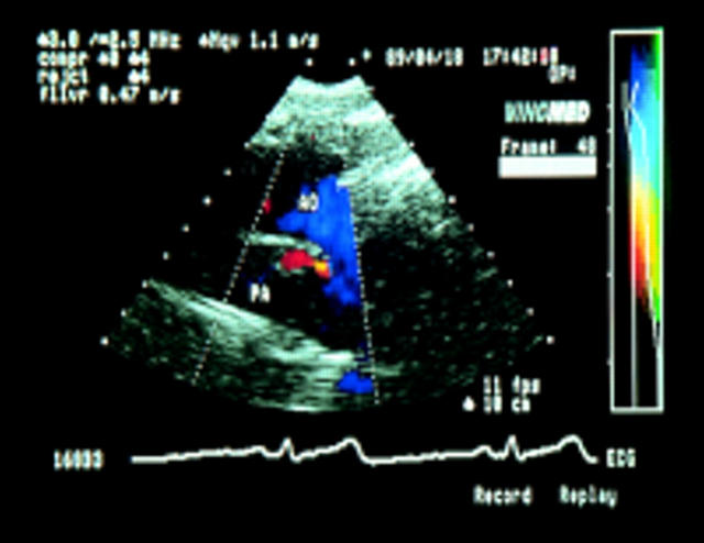
Colour Doppler demonstration of flow through the duct into the pulmonary artery in a woman with a "silent" duct. PA, pulmonary artery; AO, aorta; others as before.
Figure 13 .
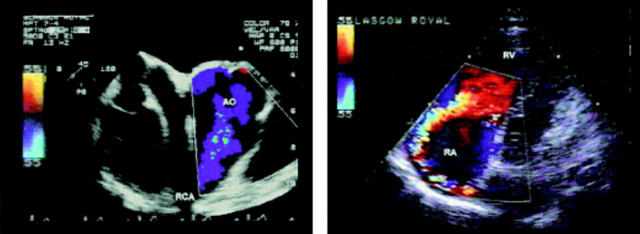
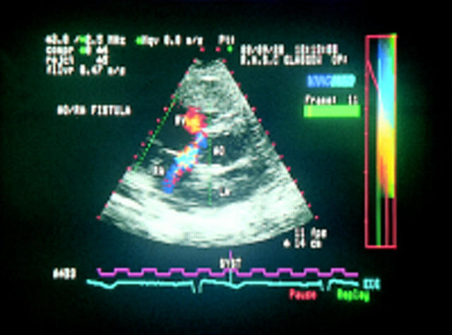
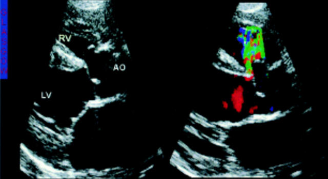
Images from a man with a fistula from the right coronary artery to the right atrium. The transoesophageal image (left) shows a very dilated right coronary artery which passes anteriorly and then sharply posteriorly. The transthoracic image (right) shows the jet flowing into the right atrium towards the right wall and then anteriorly; others as before.
Figure 14 .
Colour Doppler image in a man with a sinus of Valsalva aneurysm showing flow from the aortic root into the right atrium.
Figure 15 .
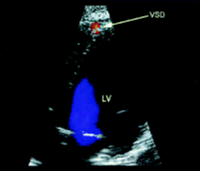
Colour Doppler image of a tiny apical ventricular septal defect (VSD). The defect cannot be shown with imaging alone but is readily identified with colour.
Figure 16 .
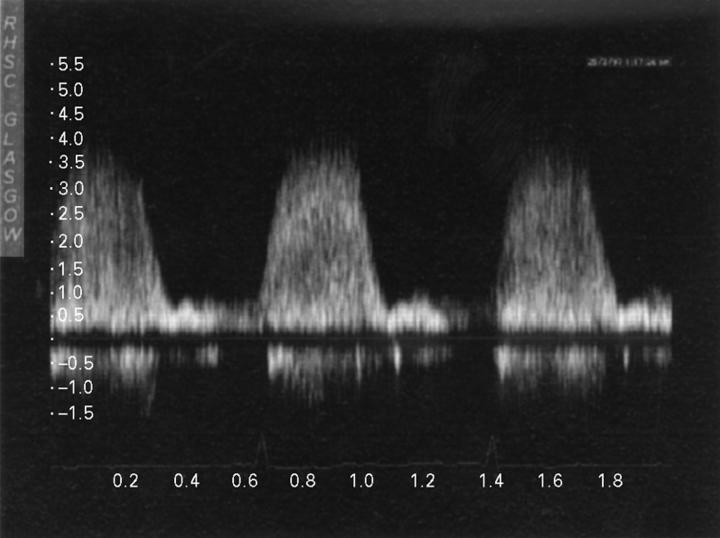
Spectral Doppler recording of flow through a small ventricular septal defect from left to right ventricle. It demonstrates a sharp rise in systole to the mid-systolic plateau and then a sharp fall at end systole. The maximum velocity of about 4 m/s represents a pressure difference between the ventricles of about 64 mm Hg.
Figure 17 .
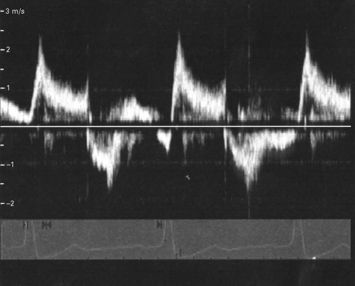
Spectral Doppler recording of flow through a ventricular septal defect in a woman with Eisenmenger's syndrome. The velocity is low (maximum 2.3 m/s) and the pattern demonstrates flow from left to right (above the line) in systole and bidirectional in diastole.
Figure 18 .
Transoesophageal study showing a mid-muscular ventricular septal defect in a man in whom it was poorly seen with transthoracic echocardiography.
Figure 19 .
Long axis views in a patient with subaortic ventricular septal defect and aortic valve prolapse. The image shows that the right coronary cusp is prolapsing into the defect and limiting its size and the colour demonstrates the residual flow through the lower part of the ventricular septal defect.
Figure 20 .
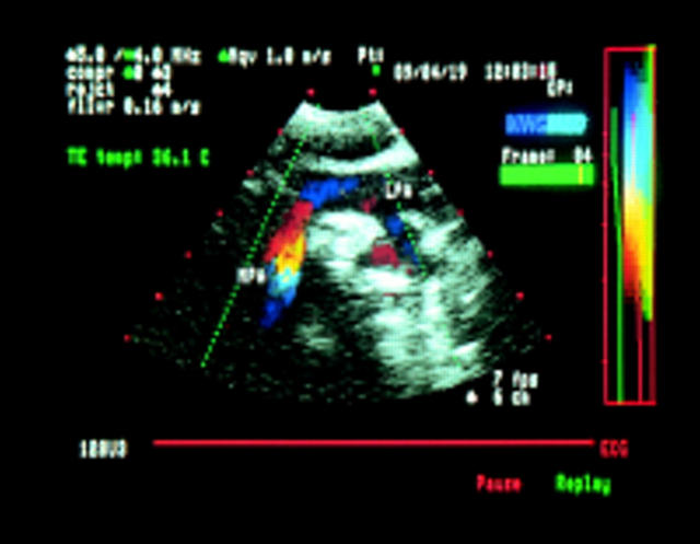
Transoesophageal image of the main pulmonary artery (MPA) and left pulmonary artery (LPA) in a patient who has undergone repair of tetralogy of Fallot. The narrowing in the MPA is indicated by the increased flow velocity on colour Doppler. The LPA and its branches are of good size with no narrowed areas.
Figure 21 .
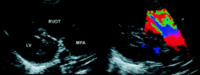
Views along the right ventricular outflow tract (RVOT) and main pulmonary artery in a patient who has undergone repair of tetralogy of Fallot. The right ventricle, outflow tract and pulmonary artery are dilated and there is jet of pulmonary regurgitation occupying the full width of the pulmonary ring.
Figure 22 .
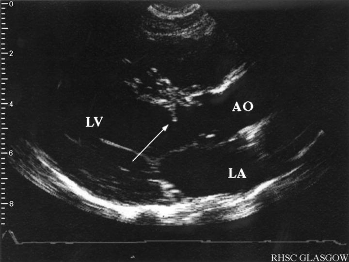
Long axis view in a patent with a subaortic membrane (arrow).
Figure 23 .
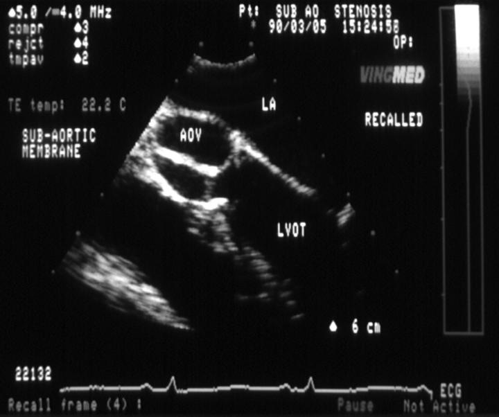
Transoesophageal echocardiography image of a subaortic membrane which is very closely related to the aortic valve and difficult to show with transthoracic imaging. AOV, aortic valve; LVOT, left ventricular outflow tract; others as before.
Figure 24 .
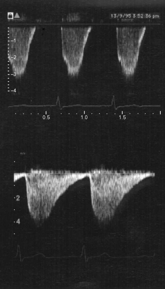
Spectral recordings of flow in the distal aortic arch from patients with mild (upper) and severe (lower) coarctation. In addition to the higher velocity of the severe obstruction the signal continues throughout diastole while in the less severe one the signal is only systolic.
Figure 25 .
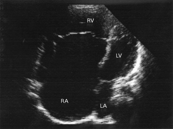
Four chamber view in a patient with Ebstein's anomaly of the tricuspid valve showing it to be apically displaced. The left ventricle (LV) and atrium (LA) are small and the right atrium (RA) large consisting of the true atrium and the "atrialised" right ventricle (RV).
Figure 26 .
Four chamber view in a patient with corrected transposition of the great arteries. The left sided valve is more apically positioned indicating a left sided right ventricle (RV) and thus a right sided left ventricle (LV).
Figure 27 .
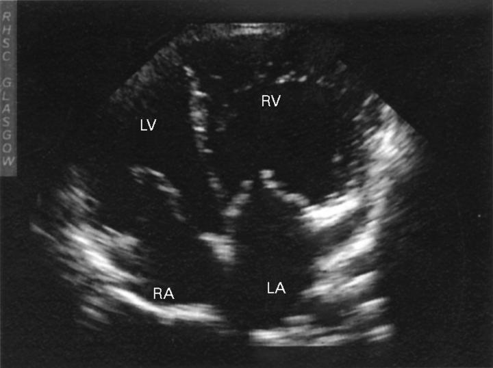
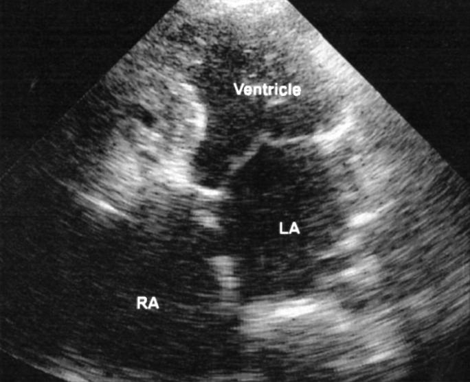
Four chamber view in a patient with tricuspid atresia and a Fontan repair. There is no apparent tricuspid valve and a single ventricular chamber. There appears to be a break in the atrial septum but this is echo dropout and none was demonstrated with colour imaging.
Figure 28 .
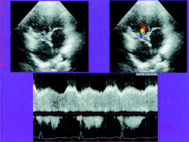
Transthoracic apical four chamber study of total cavopulmonary connection. The left image shows a double inlet ventricle and the proximity of the intra-atrial conduit to the right valve and the right shows colour flow through a surgically created fenestration and then through the atrioventricular valve to the ventricular chamber. The spectral Doppler trace of flow through the fenestration (below) shows right to left flow which is continuous and of relatively high velocity.
Figure 29 .
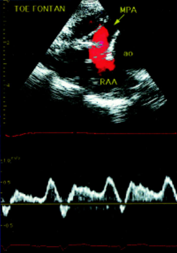
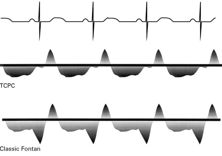
Transoesophageal study in a patient with a classic Fontan showing flow through the anastomosis and into the main pulmonary artery (MPA) on colour. The spectral signal (below) shows the typical triphasic appearance with forward flow (towards the transducer) in atrial systole followed by a brief retrograde flow in atrial relaxation and subsequent forward flow of lower velocity until atrial systole. RAA, right atrial appendage.
Figure 30 .
Diagrammatic representation of the spectral flow signals. With classic Fontan there is a distinct peak of forward flow with atrial systole, but this is lost in the total cavopulmonary connection (TCPC).
Figure 31 .
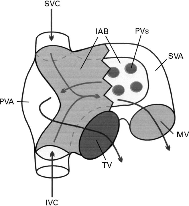
Schematic diagram demonstrating inflow redirection in the Mustard and Senning procedure. An intra-atrial baffle (IAB) is constructed from the mouth of each of the venae cavae and channelled to the left of the pulmonary veins to join and form a single channel directed to the mitral valve (MV) orifice. The pulmonary venous flow passes anterior to the inferior limb into the tricuspid valve (TV) orifice. IVC, inferior vena cava; PVA, pulmonary venous atrium; PVs, pulmonary veins; SVA, systemic venous atrium; SVC, superior vena cava.
Figure 32 .
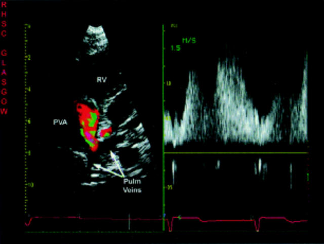
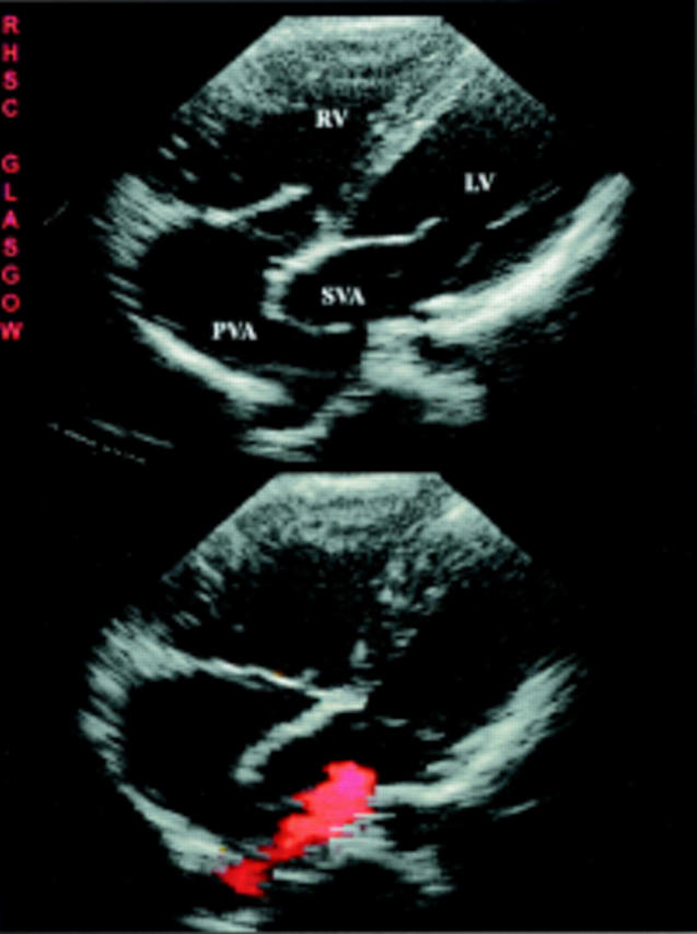
Modified subcostal transthoracic view in a Mustard correction with colour Doppler demonstrating flow acceleration through the pulmonary venous atrial anastomosis and the spectral signal showing continuous flow. PVA, pulmonary venous atrium; Pulm veins, pulmonary veins.
Figure 33 .
Parasternal four chamber views of patient with a Mustard procedure and pulmonary to systemic baffle leak. The upper image is in end diastole and shows the right ventricle (RV) to be enlarged and hypertrophied but the left ventricular (LV) size is not reduced because of a pulmonary to systemic shunt. A right sided pulmonary vein is seen draining into the pulmonary venous chamber. The lower frame is taken in systole and colour Doppler shows a low velocity shunt through the baffle leak.
Selected References
These references are in PubMed. This may not be the complete list of references from this article.
- Alboliras E. T., Porter C. B., Danielson G. K., Puga F. J., Schaff H. V., Rice M. J., Driscoll D. J. Results of the modified Fontan operation for congenital heart lesions in patients without preoperative sinus rhythm. J Am Coll Cardiol. 1985 Jul;6(1):228–233. doi: 10.1016/s0735-1097(85)80280-1. [DOI] [PubMed] [Google Scholar]
- Anderson R. H. Simplifying the understanding of congenital malformations of the heart. Int J Cardiol. 1991 Aug;32(2):131–142. doi: 10.1016/0167-5273(91)90322-g. [DOI] [PubMed] [Google Scholar]
- Ashfaq M., Houston A. B., Gnanapragasam J. P., Lilley S., Murtagh E. P. Balloon atrial septostomy under echocardiographic control: six years' experience and evaluation of the practicability of cannulation via the umbilical vein. Br Heart J. 1991 Mar;65(3):148–151. doi: 10.1136/hrt.65.3.148. [DOI] [PMC free article] [PubMed] [Google Scholar]
- Belkin R. N., Pollack B. D., Ruggiero M. L., Alas L. L., Tatini U. Comparison of transesophageal and transthoracic echocardiography with contrast and color flow Doppler in the detection of patent foramen ovale. Am Heart J. 1994 Sep;128(3):520–525. doi: 10.1016/0002-8703(94)90626-2. [DOI] [PubMed] [Google Scholar]
- Belkin R. N., Waugh R. A., Kisslo J. Interatrial shunting in atrial septal aneurysm. Am J Cardiol. 1986 Feb 1;57(4):310–312. doi: 10.1016/0002-9149(86)90909-4. [DOI] [PubMed] [Google Scholar]
- Berdjis F., Brandl D., Uhlemann F., Hausdorf G., Lange L., Weng Y., Loebe M., Alexi V., Hetzer R., Lange P. E. Erwachsene mit angeborenen Herzfehlern--klinisches Spektrum und operative Behandlung. Herz. 1996 Oct;21(5):330–336. [PubMed] [Google Scholar]
- Blakenberg F., Rhee J., Hardy C., Helton G., Higgins S. S., Higgins C. B. MRI vs echocardiography in the evaluation of the Jatene procedure. J Comput Assist Tomogr. 1994 Sep-Oct;18(5):749–754. doi: 10.1097/00004728-199409000-00013. [DOI] [PubMed] [Google Scholar]
- Bull K. The Fontan procedure: lessons from the past. Heart. 1998 Mar;79(3):213–214. doi: 10.1136/hrt.79.3.213. [DOI] [PMC free article] [PubMed] [Google Scholar]
- Canter C. E., Gutierrez F. R., Molina P., Hartmann A. F., Jr, Spray T. L. Noninvasive diagnosis of right-sided extracardiac conduit obstruction by combined magnetic resonance imaging and continuous-wave Doppler echocardiography. J Thorac Cardiovasc Surg. 1991 Apr;101(4):724–731. [PubMed] [Google Scholar]
- Chan K. C., Dickinson D. F., Wharton G. A., Gibbs J. L. Continuous wave Doppler echocardiography after surgical repair of coarctation of the aorta. Br Heart J. 1992 Aug;68(2):192–194. doi: 10.1136/hrt.68.8.192. [DOI] [PMC free article] [PubMed] [Google Scholar]
- Chen C., Kremer P., Schroeder E., Rodewald G., Bleifeld W. Usefulness of anatomic parameters derived from two-dimensional echocardiography for estimating magnitude of left to right shunt in patients with atrial septal defect. Clin Cardiol. 1987 Jun;10(6):316–321. doi: 10.1002/clc.4960100604. [DOI] [PubMed] [Google Scholar]
- Chen Y. T., Lee Y. S., Kan M. N., Chen J. S., Hu W. S., Lin W. W., Wang K. Y., Lin C. J., Chiang B. N. Transesophageal echocardiography in adults with a continuous precordial murmur. Int J Cardiol. 1992 Jul;36(1):61–68. doi: 10.1016/0167-5273(92)90109-g. [DOI] [PubMed] [Google Scholar]
- Craig B. G., Smallhorn J. F., Burrows P., Trusler G. A., Rowe R. D. Cross-sectional echocardiography in the evaluation of aortic valve prolapse associated with ventricular septal defect. Am Heart J. 1986 Oct;112(4):800–807. doi: 10.1016/0002-8703(86)90477-1. [DOI] [PubMed] [Google Scholar]
- Cromme-Dijkhuis A. H., Schasfoort-van Leeuwen M., Bink-Boekens M. T., Talsma M. The value of 2-D Doppler echocardiography in the evaluation of asymptomatic patients with Mustard operation for transposition of the great arteries. Eur Heart J. 1991 Dec;12(12):1308–1310. doi: 10.1093/eurheartj/12.12.1308. [DOI] [PubMed] [Google Scholar]
- Cross S. J., Evans S. A., Thomson L. F., Lee H. S., Jennings K. P., Shields T. G. Safety of subaqua diving with a patent foramen ovale. BMJ. 1992 Feb 22;304(6825):481–482. doi: 10.1136/bmj.304.6825.481. [DOI] [PMC free article] [PubMed] [Google Scholar]
- Davis R. H., Feigenbaum H., Chang S., Konecke L. L., Dillon J. C. Echocardiographic manifestations of discrete subaortic stenosis. Am J Cardiol. 1974 Feb;33(2):277–280. doi: 10.1016/0002-9149(74)90289-6. [DOI] [PubMed] [Google Scholar]
- DiSessa T. G., Child J. S., Perloff J. K., Wu L., Williams R. G., Laks H., Friedman W. F. Systemic venous and pulmonary arterial flow patterns after Fontan's procedure for tricuspid atresia or single ventricle. Circulation. 1984 Nov;70(5):898–902. doi: 10.1161/01.cir.70.5.898. [DOI] [PubMed] [Google Scholar]
- DiSessa T. G., Hagan A. D., Isabel-Jones J. B., Ti C. C., Mercier J. C., Friedman W. F. Two-dimensional echocardiographic evaluation of discrete subaortic stenosis from the apical long axis view. Am Heart J. 1981 Jun;101(6):774–782. doi: 10.1016/0002-8703(81)90615-3. [DOI] [PubMed] [Google Scholar]
- Diamond M. A., Dillon J. C., Haine C. L., Chang S., Feigenbaum H. Echocardiographic features of atrial septal defect. Circulation. 1971 Jan;43(1):129–135. doi: 10.1161/01.cir.43.1.129. [DOI] [PubMed] [Google Scholar]
- Duncan W. J., Ninomiya K., Cook D. H., Rowe R. D. Noninvasive diagnosis of neonatal coarctation and associated anomalies using two-dimensional echocardiography. Am Heart J. 1983 Jul;106(1 Pt 1):63–69. doi: 10.1016/0002-8703(83)90441-6. [DOI] [PubMed] [Google Scholar]
- Eichhorn P., Sütsch G., Jenni R. Echokardiographisch neu entdeckte kongenitale Vitien und Anomalien bei Adoleszenten und Erwachsenen. Schweiz Med Wochenschr. 1990 Nov 10;120(45):1697–1700. [PubMed] [Google Scholar]
- Eichhorn P., Vogt P., Ritter M., Widmer V., Jenni R. Anomalien des Vorhofseptums bei Erwachsenen: Art, Häufigkeit und klinische Relevanz. Schweiz Med Wochenschr. 1995 Jul 11;125(27-28):1336–1341. [PubMed] [Google Scholar]
- Engvall J., Sjöqvist L., Nylander E., Thuomas K. A., Wranne B. Biplane transoesophageal echocardiography, transthoracic Doppler, and magnetic resonance imaging in the assessment of coarctation of the aorta. Eur Heart J. 1995 Oct;16(10):1399–1409. doi: 10.1093/oxfordjournals.eurheartj.a060748. [DOI] [PubMed] [Google Scholar]
- Fontan F., Baudet E. Surgical repair of tricuspid atresia. Thorax. 1971 May;26(3):240–248. doi: 10.1136/thx.26.3.240. [DOI] [PMC free article] [PubMed] [Google Scholar]
- Fontan F., Kirklin J. W., Fernandez G., Costa F., Naftel D. C., Tritto F., Blackstone E. H. Outcome after a "perfect" Fontan operation. Circulation. 1990 May;81(5):1520–1536. doi: 10.1161/01.cir.81.5.1520. [DOI] [PubMed] [Google Scholar]
- Frommelt P. C., Snider A. R., Meliones J. N., Vermilion R. P. Doppler assessment of pulmonary artery flow patterns and ventricular function after the Fontan operation. Am J Cardiol. 1991 Nov 1;68(11):1211–1215. doi: 10.1016/0002-9149(91)90195-q. [DOI] [PubMed] [Google Scholar]
- Fyfe D. A., Kline C. H., Sade R. M., Gillette P. C. Transesophageal echocardiography detects thrombus formation not identified by transthoracic echocardiography after the Fontan operation. J Am Coll Cardiol. 1991 Dec;18(7):1733–1737. doi: 10.1016/0735-1097(91)90512-8. [DOI] [PubMed] [Google Scholar]
- Gibbs J. L., Qureshi S. A., Grieve L., Webb C., Smith R. R., Yacoub M. H. Doppler echocardiography after anatomical correction of transposition of the great arteries. Br Heart J. 1986 Jul;56(1):67–72. doi: 10.1136/hrt.56.1.67. [DOI] [PMC free article] [PubMed] [Google Scholar]
- Gnanapragasam J. P., Houston A. B., Doig W. B., Jamieson M. P., Pollock J. C. Transoesophageal echocardiographic assessment of fixed subaortic obstruction in children. Br Heart J. 1991 Oct;66(4):281–284. doi: 10.1136/hrt.66.4.281. [DOI] [PMC free article] [PubMed] [Google Scholar]
- Gnanapragasam J. P., Houston A. B., Northridge D. B., Jamieson M. P., Pollock J. C. Transoesophageal echocardiographic assessment of primum, secundum and sinus venosus atrial septal defects. Int J Cardiol. 1991 May;31(2):167–174. doi: 10.1016/0167-5273(91)90212-8. [DOI] [PubMed] [Google Scholar]
- Hagen P. T., Scholz D. G., Edwards W. D. Incidence and size of patent foramen ovale during the first 10 decades of life: an autopsy study of 965 normal hearts. Mayo Clin Proc. 1984 Jan;59(1):17–20. doi: 10.1016/s0025-6196(12)60336-x. [DOI] [PubMed] [Google Scholar]
- Hagler D. J., Edwards W. D., Seward J. B., Tajik A. J. Standardized nomenclature of the ventricular septum and ventricular septal defects, with applications for two-dimensional echocardiography. Mayo Clin Proc. 1985 Nov;60(11):741–752. doi: 10.1016/s0025-6196(12)60416-9. [DOI] [PubMed] [Google Scholar]
- Hagler D. J., Tajik A. J., Seward J. B., Mair D. D., Ritter D. G. Real-time wide-angle sector echocardiography: atrioventricular canal defects. Circulation. 1979 Jan;59(1):140–150. doi: 10.1161/01.cir.59.1.140. [DOI] [PubMed] [Google Scholar]
- Harrison D. A., Liu P., Walters J. E., Goodman J. M., Siu S. C., Webb G. D., Williams W. G., McLaughlin P. R. Cardiopulmonary function in adult patients late after Fontan repair. J Am Coll Cardiol. 1995 Oct;26(4):1016–1021. doi: 10.1016/0735-1097(95)00242-7. [DOI] [PubMed] [Google Scholar]
- Hoffmann R., Lambertz H., Flachskampf F. A., Hanrath P. Transoesophageal echocardiography in the diagnosis of cor triatriatum; incremental value of colour Doppler. Eur Heart J. 1992 Mar;13(3):418–420. doi: 10.1093/oxfordjournals.eurheartj.a060184. [DOI] [PubMed] [Google Scholar]
- Hopkins W. E., Waggoner A. D., Davila-Roman V., Perez J. E. Two-dimensional Doppler color flow imaging in adults with L-transposition of the great arteries. Echocardiography. 1993 Nov;10(6):611–617. doi: 10.1111/j.1540-8175.1993.tb00078.x. [DOI] [PubMed] [Google Scholar]
- Houston A. B., Lim M. K., Doig W. B., Reid J. M., Coleman E. N. Doppler assessment of the interventricular pressure drop in patients with ventricular septal defects. Br Heart J. 1988 Jul;60(1):50–56. doi: 10.1136/hrt.60.1.50. [DOI] [PMC free article] [PubMed] [Google Scholar]
- Houston A. B., Simpson I. A., Pollock J. C., Jamieson M. P., Doig W. B., Coleman E. N. Doppler ultrasound in the assessment of severity of coarctation of the aorta and interruption of the aortic arch. Br Heart J. 1987 Jan;57(1):38–43. doi: 10.1136/hrt.57.1.38. [DOI] [PMC free article] [PubMed] [Google Scholar]
- Huhta J. C., Seward J. B., Tajik A. J., Hagler D. J., Edwards W. D. Two-dimensional echocardiographic spectrum of univentricular atrioventricular connection. J Am Coll Cardiol. 1985 Jan;5(1):149–157. doi: 10.1016/s0735-1097(85)80098-x. [DOI] [PubMed] [Google Scholar]
- Huhta J. C., Smallhorn J. F., Macartney F. J. Two dimensional echocardiographic diagnosis of situs. Br Heart J. 1982 Aug;48(2):97–108. doi: 10.1136/hrt.48.2.97. [DOI] [PMC free article] [PubMed] [Google Scholar]
- Hutchison Stuart J., Rosin Benjamin L., Curry Susan, Chandraratna P. Anthony N. Transesophageal Echocardiographic Assessment of Lesions of the Right Ventricular Outflow Tract and Pulmonic Valve. Echocardiography. 1996 Jan;13(1):21–34. doi: 10.1111/j.1540-8175.1996.tb00864.x. [DOI] [PubMed] [Google Scholar]
- Jobic Y., Slama M., Tribouilloy C., Lan Cheong Wah L., Choquet D., Boschat J., Penther P., Lesbre J. P. Doppler echocardiographic evaluation of valve regurgitation in healthy volunteers. Br Heart J. 1993 Feb;69(2):109–113. doi: 10.1136/hrt.69.2.109. [DOI] [PMC free article] [PubMed] [Google Scholar]
- Jonsson H., Ivert T., Brodin L. A. Echocardiographic findings in 83 patients 13-26 years after intracardiac repair of tetralogy of Fallot. Eur Heart J. 1995 Sep;16(9):1255–1263. doi: 10.1093/oxfordjournals.eurheartj.a061083. [DOI] [PubMed] [Google Scholar]
- Kaulitz R., Stümper O. F., Geuskens R., Sreeram N., Elzenga N. J., Chan C. K., Burns J. E., Godman M. J., Hess J., Sutherland G. R. Comparative values of the precordial and transesophageal approaches in the echocardiographic evaluation of atrial baffle function after an atrial correction procedure. J Am Coll Cardiol. 1990 Sep;16(3):686–694. doi: 10.1016/0735-1097(90)90361-r. [DOI] [PubMed] [Google Scholar]
- Ke W. L., Wang N. K., Lin Y. M., Shen C. T., Chen C. C. Right coronary artery fistula into right atrium: diagnosis by color Doppler echocardiography. Am Heart J. 1988 Sep;116(3):886–889. doi: 10.1016/0002-8703(88)90358-4. [DOI] [PubMed] [Google Scholar]
- Kronzon I., Tunick P. A., Freedberg R. S., Trehan N., Rosenzweig B. P., Schwinger M. E. Transesophageal echocardiography is superior to transthoracic echocardiography in the diagnosis of sinus venosus atrial septal defect. J Am Coll Cardiol. 1991 Feb;17(2):537–542. doi: 10.1016/s0735-1097(10)80128-7. [DOI] [PubMed] [Google Scholar]
- Lim M. K., Houston A. B., Doig W. B., Lilley S., Murtagh E. P. Variability of the Doppler gradient in pulmonary valve stenosis before and after balloon dilatation. Br Heart J. 1989 Sep;62(3):212–216. doi: 10.1136/hrt.62.3.212. [DOI] [PMC free article] [PubMed] [Google Scholar]
- Lima C. O., Sahn D. J., Valdes-Cruz L. M., Goldberg S. J., Barron J. V., Allen H. D., Grenadier E. Noninvasive prediction of transvalvular pressure gradient in patients with pulmonary stenosis by quantitative two-dimensional echocardiographic Doppler studies. Circulation. 1983 Apr;67(4):866–871. doi: 10.1161/01.cir.67.4.866. [DOI] [PubMed] [Google Scholar]
- Marcella C. P., Johnson L. E. Right parasternal imaging: an underutilized echocardiographic technique. J Am Soc Echocardiogr. 1993 Jul-Aug;6(4):453–466. doi: 10.1016/s0894-7317(14)80245-9. [DOI] [PubMed] [Google Scholar]
- Mascarenhas E., Javier R. P., Samet P. Partial anomalous pulmonary venous connection and drainage. Am J Cardiol. 1973 Apr;31(4):512–518. doi: 10.1016/0002-9149(73)90304-4. [DOI] [PubMed] [Google Scholar]
- Minich L. L., Snider A. R. Echocardiographic guidance during placement of the buttoned double-disk device for atrial septal defect closure. Echocardiography. 1993 Nov;10(6):567–572. doi: 10.1111/j.1540-8175.1993.tb00072.x. [DOI] [PubMed] [Google Scholar]
- Mortera C., Rissech M., Payola M., Miro C., Perich R. Cross sectional subcostal echocardiography: atrioventricular septal defects and the short axis cut. Br Heart J. 1987 Sep;58(3):267–273. doi: 10.1136/hrt.58.3.267. [DOI] [PMC free article] [PubMed] [Google Scholar]
- Mulhern K. M., Skorton D. J. Echocardiographic evaluation of isolated pulmonary valve disease in adolescents and adults. Echocardiography. 1993 Sep;10(5):533–543. doi: 10.1111/j.1540-8175.1993.tb00068.x. [DOI] [PubMed] [Google Scholar]
- Mügge A., Daniel W. G., Wolpers H. G., Klöpper J. W., Lichtlen P. R. Improved visualization of discrete subvalvular aortic stenosis by transesophageal color-coded Doppler echocardiography. Am Heart J. 1989 Feb;117(2):474–475. doi: 10.1016/0002-8703(89)90795-3. [DOI] [PubMed] [Google Scholar]
- Penny D. J., Redington A. N. Doppler echocardiographic evaluation of pulmonary blood flow after the Fontan operation: the role of the lungs. Br Heart J. 1991 Nov;66(5):372–374. doi: 10.1136/hrt.66.5.372. [DOI] [PMC free article] [PubMed] [Google Scholar]
- Qureshi S. A., Richheimer R., McKay R., Arnold R. Doppler echocardiographic evaluation of pulmonary artery flow after modified Fontan operation: importance of atrial contraction. Br Heart J. 1990 Oct;64(4):272–276. doi: 10.1136/hrt.64.4.272. [DOI] [PMC free article] [PubMed] [Google Scholar]
- Rao P. S., Carey P. Doppler ultrasound in the prediction of pressure gradients across aortic coarctation. Am Heart J. 1989 Aug;118(2):299–307. doi: 10.1016/0002-8703(89)90189-0. [DOI] [PubMed] [Google Scholar]
- Rebergen S. A., Ottenkamp J., Doornbos J., van der Wall E. E., Chin J. G., de Roos A. Postoperative pulmonary flow dynamics after Fontan surgery: assessment with nuclear magnetic resonance velocity mapping. J Am Coll Cardiol. 1993 Jan;21(1):123–131. doi: 10.1016/0735-1097(93)90726-h. [DOI] [PubMed] [Google Scholar]
- Redington A. N., Rigby M. L., Oldershaw P., Gibson D. G., Shinebourne E. A. Right ventricular function 10 years after the Mustard operation for transposition of the great arteries: analysis of size, shape, and wall motion. Br Heart J. 1989 Dec;62(6):455–461. doi: 10.1136/hrt.62.6.455. [DOI] [PMC free article] [PubMed] [Google Scholar]
- Rees S., Somerville J., Ward C., Martinez J., Mohiaddin R. H., Underwood R., Longmore D. B. Coarctation of the aorta: MR imaging in late postoperative assessment. Radiology. 1989 Nov;173(2):499–502. doi: 10.1148/radiology.173.2.2798882. [DOI] [PubMed] [Google Scholar]
- Rodgers D. M., Wolf N. M., Barrett M. J., Zuckerman G. L., Meister S. G. Two-dimensional echocardiographic features of coronary arteriovenous fistula. Am Heart J. 1982 Oct;104(4 Pt 1):872–874. [PubMed] [Google Scholar]
- Sagin-Saylam G., Somerville J. Palliative Mustard operation for transposition of the great arteries: late results after 15-20 years. Heart. 1996 Jan;75(1):72–77. doi: 10.1136/hrt.75.1.72. [DOI] [PMC free article] [PubMed] [Google Scholar]
- Salzer-Muhar U., Proll E., Marx M., Salzer H. R., Wimmer M. Two-dimensional and Doppler echocardiographic follow-up after the arterial switch operation for transposition of the great arteries. Thorac Cardiovasc Surg. 1991 Dec;39 (Suppl 2):180–184. doi: 10.1055/s-2007-1020015. [DOI] [PubMed] [Google Scholar]
- Sapin P., Frantz E., Jain A., Nichols T. C., Dehmer G. J. Coronary artery fistula: an abnormality affecting all age groups. Medicine (Baltimore) 1990 Mar;69(2):101–113. [PubMed] [Google Scholar]
- Shiina A., Seward J. B., Edwards W. D., Hagler D. J., Tajik A. J. Two-dimensional echocardiographic spectrum of Ebstein's anomaly: detailed anatomic assessment. J Am Coll Cardiol. 1984 Feb;3(2 Pt 1):356–370. doi: 10.1016/s0735-1097(84)80020-0. [DOI] [PubMed] [Google Scholar]
- Shub C., Dimopoulos I. N., Seward J. B., Callahan J. A., Tancredi R. G., Schattenberg T. T., Reeder G. S., Hagler D. J., Tajik A. J. Sensitivity of two-dimensional echocardiography in the direct visualization of atrial septal defect utilizing the subcostal approach: experience with 154 patients. J Am Coll Cardiol. 1983 Jul;2(1):127–135. doi: 10.1016/s0735-1097(83)80385-4. [DOI] [PubMed] [Google Scholar]
- Shyu K. G., Chen J. J., Huang Z. S., Hwang J. J., Lee T. K., Kuan P., Lien W. P. Role of transesophageal echocardiography in the diagnostic assessment of cardiac sources of embolism in patients with acute ischemic stroke. Cardiology. 1994;85(1):53–60. doi: 10.1159/000176646. [DOI] [PubMed] [Google Scholar]
- Shyu K. G., Lai L. P., Lin S. C., Chang H., Chen J. J. Diagnostic accuracy of transesophageal echocardiography for detecting patent ductus arteriosus in adolescents and adults. Chest. 1995 Nov;108(5):1201–1205. doi: 10.1378/chest.108.5.1201. [DOI] [PubMed] [Google Scholar]
- Silverman N. H., Payot M., Stanger P., Rudolph A. M. The echocardiographic profile of patients after Mustard's operation. Circulation. 1978 Dec;58(6):1083–1093. doi: 10.1161/01.cir.58.6.1083. [DOI] [PubMed] [Google Scholar]
- Silverman N. H., Snider A. R., Coló J., Ebert P. A., Turley K. Superior vena caval obstruction after Mustard's operation: detection by two-dimensional contrast echocardiography. Circulation. 1981 Aug;64(2):392–396. doi: 10.1161/01.cir.64.2.392. [DOI] [PubMed] [Google Scholar]
- Simpson I. A., de Belder M. A., Kenny A., Martin M., Nihoyannopoulos P. How to quantitate valve regurgitation by echo Doppler techniques. British Society of Echocardiography. Br Heart J. 1995 May;73(5 Suppl 2):1–9. doi: 10.1136/hrt.73.5_suppl_2.1. [DOI] [PMC free article] [PubMed] [Google Scholar]
- Smallhorn J. F., Huhta J. C., Anderson R. H., Macartney F. J. Suprasternal cross-sectional echocardiography in assessment of patient ducts arteriosus. Br Heart J. 1982 Oct;48(4):321–330. doi: 10.1136/hrt.48.4.321. [DOI] [PMC free article] [PubMed] [Google Scholar]
- Smallhorn J. F., Tommasini G., Anderson R. H., Macartney F. J. Assessment of atrioventricular septal defects by two dimensional echocardiography. Br Heart J. 1982 Feb;47(2):109–121. doi: 10.1136/hrt.47.2.109. [DOI] [PMC free article] [PubMed] [Google Scholar]
- Sreeram N., Sutherland G. R., Geuskens R., Stümper O. F., Taams M., Gussenhoven E. J., Hess J., Roelandt J. R. The role of transoesophageal echocardiography in adolescents and adults with congenital heart defects. Eur Heart J. 1991 Feb;12(2):231–240. doi: 10.1093/oxfordjournals.eurheartj.a059874. [DOI] [PubMed] [Google Scholar]
- Stümper O., Sutherland G. R., Geuskens R., Roelandt J. R., Bos E., Hess J. Transesophageal echocardiography in evaluation and management after a Fontan procedure. J Am Coll Cardiol. 1991 Apr;17(5):1152–1160. doi: 10.1016/0735-1097(91)90847-3. [DOI] [PubMed] [Google Scholar]
- Sutherland G. R., Godman M. J., Smallhorn J. F., Guiterras P., Anderson R. H., Hunter S. Ventricular septal defects. Two dimensional echocardiographic and morphological correlations. Br Heart J. 1982 Apr;47(4):316–328. doi: 10.1136/hrt.47.4.316. [DOI] [PMC free article] [PubMed] [Google Scholar]
- Sutherland G. R., Stümper O. F. Transoesophageal echocardiography in congenital heart disease. Acta Paediatr Suppl. 1995 Aug;410:15–22. doi: 10.1111/j.1651-2227.1995.tb13840.x. [DOI] [PubMed] [Google Scholar]
- Swensson R. E., Valdes-Cruz L. M., Sahn D. J., Sherman F. S., Chung K. J., Scagnelli S., Hagen-Ansert S. Real-time Doppler color flow mapping for detection of patent ductus arteriosus. J Am Coll Cardiol. 1986 Nov;8(5):1105–1112. doi: 10.1016/s0735-1097(86)80388-6. [DOI] [PubMed] [Google Scholar]
- Van Hare G. F., Silverman N. H. Contrast two-dimensional echocardiography in congenital heart disease: techniques, indications and clinical utility. J Am Coll Cardiol. 1989 Mar 1;13(3):673–686. doi: 10.1016/0735-1097(89)90610-4. [DOI] [PubMed] [Google Scholar]
- Vick G. W., 3rd, Murphy D. J., Jr, Ludomirsky A., Morrow W. R., Morriss M. J., Danford D. A., Huhta J. C. Pulmonary venous and systemic ventricular inflow obstruction in patients with congenital heart disease: detection by combined two-dimensional and Doppler echocardiography. J Am Coll Cardiol. 1987 Mar;9(3):580–587. doi: 10.1016/s0735-1097(87)80051-7. [DOI] [PubMed] [Google Scholar]
- Wilson N. J., Neutze J. M., Rutland M. D., Ramage M. C. Transthoracic echocardiography for right ventricular function late after the Mustard operation. Am Heart J. 1996 Feb;131(2):360–367. doi: 10.1016/s0002-8703(96)90367-1. [DOI] [PubMed] [Google Scholar]
- de Leval M. R., Kilner P., Gewillig M., Bull C. Total cavopulmonary connection: a logical alternative to atriopulmonary connection for complex Fontan operations. Experimental studies and early clinical experience. J Thorac Cardiovasc Surg. 1988 Nov;96(5):682–695. [PubMed] [Google Scholar]


